Copyright
©2007 Baishideng Publishing Group Co.
World J Gastroenterol. Aug 7, 2007; 13(29): 3925-3931
Published online Aug 7, 2007. doi: 10.3748/wjg.v13.i29.3925
Published online Aug 7, 2007. doi: 10.3748/wjg.v13.i29.3925
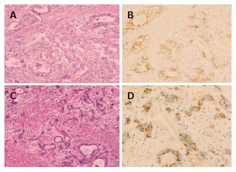
Figure 1 E-cadherin expression.
A and B: an E-cadherin-positive case with membranous pattern; C and D: an E-cadherin-positive case with cytoplasmic pattern. E-cadherin expression is observed in cytoplasm but not in nuclei of carcinoma cells (D). A and C: hematoxylin and eosin staining; B and D: Immunohistochemical staining.
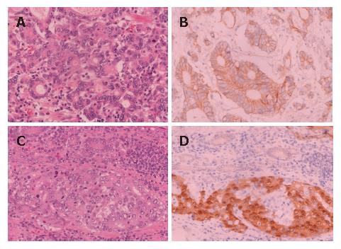
Figure 2 Beta-catenin expression.
A and B: A beta-catenin-positive case with membranous pattern; C and D: Beta-catenin expression is observed in both nuclei and cytoplasm of carcinoma cells (D). A and C: hematoxylin and eosin staining; B and D: Immunohistochemical staining.
- Citation: Koriyama C, Akiba S, Itoh T, Sueyoshi K, Minakami Y, Corvalan A, Yonezawa S, Eizuru Y. E-cadherin and beta-catenin expression in Epstein-Barr virus-associated gastric carcinoma and their prognostic significance. World J Gastroenterol 2007; 13(29): 3925-3931
- URL: https://www.wjgnet.com/1007-9327/full/v13/i29/3925.htm
- DOI: https://dx.doi.org/10.3748/wjg.v13.i29.3925


