Copyright
©2007 Baishideng Publishing Group Inc.
World J Gastroenterol. Jul 14, 2007; 13(26): 3634-3637
Published online Jul 14, 2007. doi: 10.3748/wjg.v13.i26.3634
Published online Jul 14, 2007. doi: 10.3748/wjg.v13.i26.3634
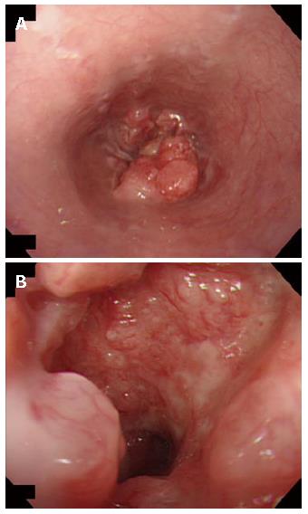
Figure 1 A: Preoperative endoscopy showing a protruding tumor of the lower esophagus; B: Close endoscopic view of the esophageal tumor with ulceration.
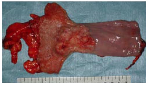
Figure 2 The resected specimen of esophagus having a protruding tumor with ulceration.
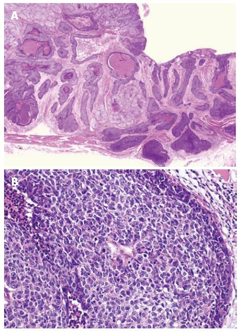
Figure 3 Histological finding of basaloid-squamous carcinomas (HE).
A: Lower magnification; B: Higher magnification.
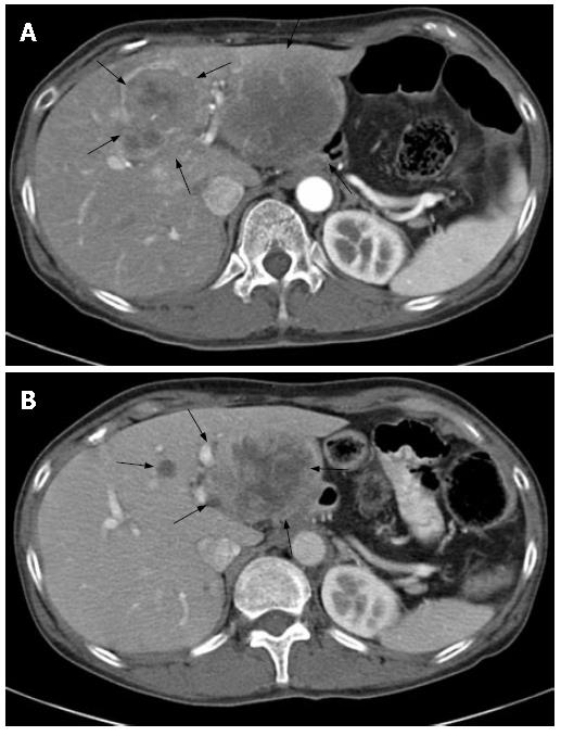
Figure 4 Abdominal computed tomography (CT) images.
A: CT image on admission revealing liver metastasis; B: CT image after 3 courses of 5FU/CDDP chemotherapy showing tumor regression (55%).
- Citation: Shibata Y, Baba E, Ariyama H, Miki R, Ogami N, Arita S, Qin B, Kusaba H, Mitsugi K, Noshiro H, Yao T, Nakano S. Metastatic basaloid-squamous cell carcinoma of the esophagus treated by 5-fluorouracil and cisplatin. World J Gastroenterol 2007; 13(26): 3634-3637
- URL: https://www.wjgnet.com/1007-9327/full/v13/i26/3634.htm
- DOI: https://dx.doi.org/10.3748/wjg.v13.i26.3634


