Copyright
©2007 Baishideng Publishing Group Inc.
World J Gastroenterol. Jul 7, 2007; 13(25): 3523-3525
Published online Jul 7, 2007. doi: 10.3748/wjg.v13.i25.3523
Published online Jul 7, 2007. doi: 10.3748/wjg.v13.i25.3523
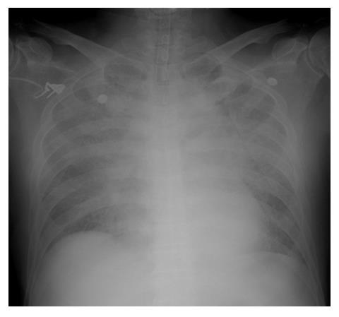
Figure 1 Chest PA.
Chest radiography shows a massive alveolar infiltrate and pleural effusion in the both lung fields.
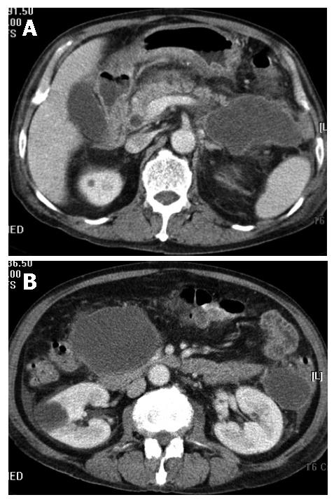
Figure 2 Abdominal CT scan.
A: CT shows a cystic lesion in the pancreatic tail and peripheral fat infiltration; B: CT shows two cystic lesions in the head and the tail.
- Citation: Yi SY, Tae JH. Pancreatic abscess following scrub typhus associated with multiorgan failure. World J Gastroenterol 2007; 13(25): 3523-3525
- URL: https://www.wjgnet.com/1007-9327/full/v13/i25/3523.htm
- DOI: https://dx.doi.org/10.3748/wjg.v13.i25.3523


