Copyright
©2007 Baishideng Publishing Group Co.
World J Gastroenterol. Jun 28, 2007; 13(24): 3392-3395
Published online Jun 28, 2007. doi: 10.3748/wjg.v13.i24.3392
Published online Jun 28, 2007. doi: 10.3748/wjg.v13.i24.3392
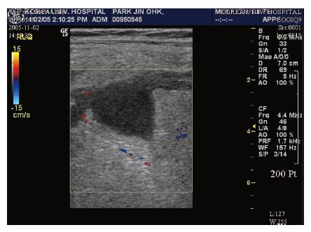
Figure 1 Homo-genous hypovascular hypo-echoic mass lesions in the mese-ntery on ultrasonography.
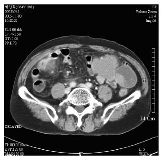
Figure 2 Lesions adjacent to small bowel loop in left abdomen and ill defined low density cystic change on computed tomography.
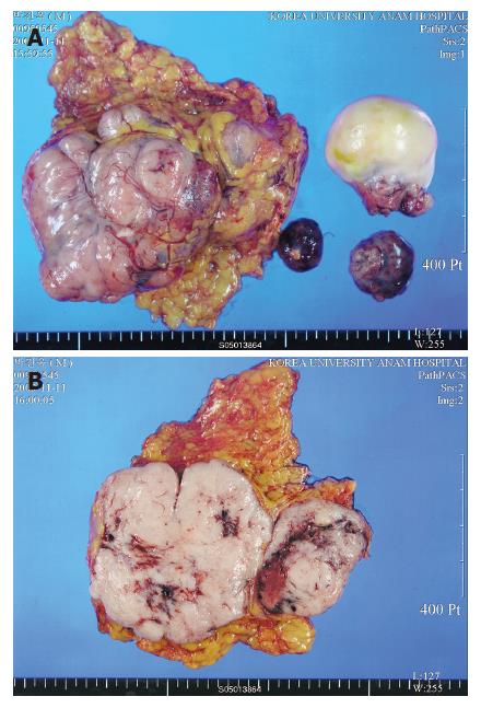
Figure 3 A: Gross findings of the specimen: two irregular fragments of omental tissue and several irregular fragments of soft tissue masses; B: Diffuse pale brownish flesh-like appearance with multiple hemorrhagic necrosis on cut surface.
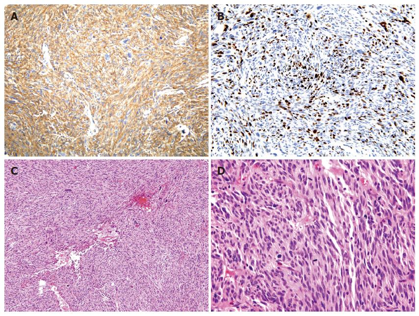
Figure 4 A: The spindle cells showed cytoplasmic positive reactivity for CD 117 (c-kit) on immunohistochemical findings; B: Ki-67 labeling index was about 30%-40% on immuno-histochemical findings; C: The tumor was composed of spindle cells showing a less developed fascicular pattern on histologic findings (x 100); D: The tumor had a high mitotic activity, about 115/50 HPFs mitotic counts on histologic findings (x 400).
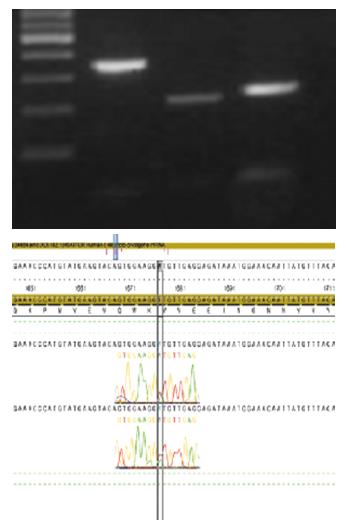
Figure 5 There was a heterozygote on exon 11, showing GTT (val.
) to GWT (val. + asp.) transition at codon 559 in c-kit mutation.
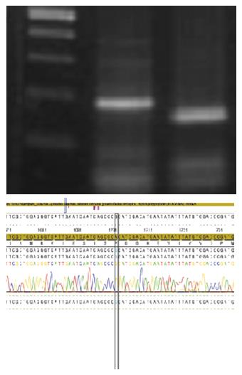
Figure 6 There was a polymorphism on exon 12, showing CAA (pro.
) to CCG (pro.) transition at codon 567 in mutation of PDGFRA.
- Citation: Kim JH, Boo YJ, Jung CW, Park SS, Kim SJ, Mok YJ, Kim SD, Chae YS, Kim CS. Multiple malignant extragastrointestinal stromal tumors of the greater omentum and results of immunohistochemistry and mutation analysis: A case report. World J Gastroenterol 2007; 13(24): 3392-3395
- URL: https://www.wjgnet.com/1007-9327/full/v13/i24/3392.htm
- DOI: https://dx.doi.org/10.3748/wjg.v13.i24.3392


