Copyright
©2007 Baishideng Publishing Group Co.
World J Gastroenterol. Jun 28, 2007; 13(24): 3388-3391
Published online Jun 28, 2007. doi: 10.3748/wjg.v13.i24.3388
Published online Jun 28, 2007. doi: 10.3748/wjg.v13.i24.3388
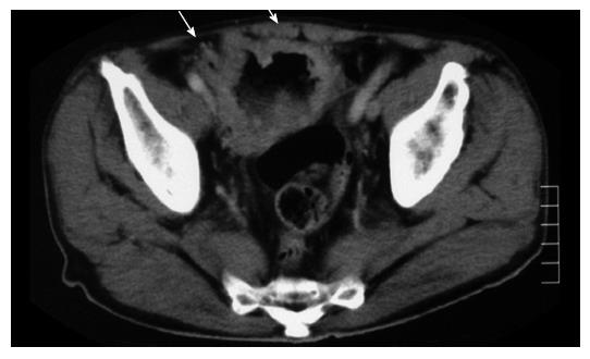
Figure 1 Computed tomography, showing localized dilation of the small intestine (arrows) in the pelvis.
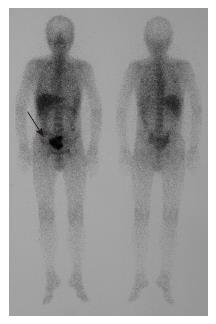
Figure 2 Gallium-67 scintigraphy, showing collection in the pelvis (arrow).
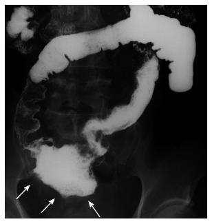
Figure 3 Double-contrast barium study of the small intestine, showing a papillary tumor in the dilated ileum (arrows).
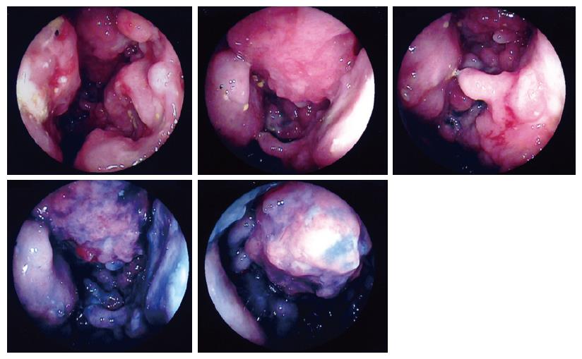
Figure 4 Double-balloon entero-doscopic examination, showing a papillary tumor at 100 cm proximal to the ileocecal valve.
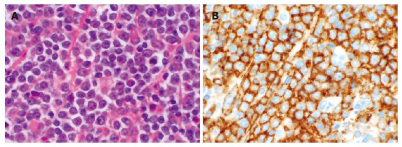
Figure 5 A: Histological findings demonstrated large atypical lymphoid lymphocytes, (HE, x 400); B: Immuno-histochemical analysis showed that these lymphocytes were positive for CD3.
(CD3, x 400).
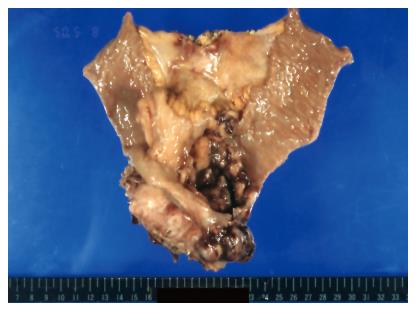
Figure 6 The resected specimen of ileum, showing an elevated lesion (110 mm × 85 mm × 50 mm) with central ulceration.
- Citation: Beppu K, Osada T, Nagahara A, Sakamoto N, Shibuya T, Kawabe M, Terai T, Ohkusa T, Ogihara T, Sato N, Kamano T, Hayashida Y, Watanabe S. Malignant lymphoma in the ileum diagnosed by double-balloon enteroscopy. World J Gastroenterol 2007; 13(24): 3388-3391
- URL: https://www.wjgnet.com/1007-9327/full/v13/i24/3388.htm
- DOI: https://dx.doi.org/10.3748/wjg.v13.i24.3388


