Copyright
©2007 Baishideng Publishing Group Co.
World J Gastroenterol. Jun 28, 2007; 13(24): 3279-3287
Published online Jun 28, 2007. doi: 10.3748/wjg.v13.i24.3279
Published online Jun 28, 2007. doi: 10.3748/wjg.v13.i24.3279
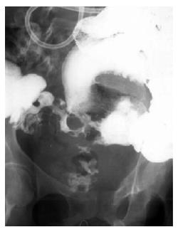
Figure 1 Small bowel enteroclysis.
Evidence of narrowing and alterations in the terminal ileum and presence of an entero-vesical fistula.
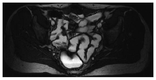
Figure 2 Magnetic resonance, axial T2-weighted fast spin-echo.
Mean age female patient with recurrent abdominal pain. This image shows homogeneus small bowel loops distention and thin wall, thus negative for Crohn's disease.
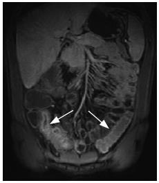
Figure 3 Magnetic resonance, coronal 3D T1-weighted gradient-echo, after intravenous administration of gadolinium contrast agent.
Young male patient with known Crohn's disease, recurrent abdominal pain in MR staging of the disease before therapy. The straight white arrow shows the last segment of small bowel with thickened wall and typical selective submucosal enhancement in the arterial phase. The curved white arrow shows a normal intestinal segment with thin wall and no contrast enhancement.
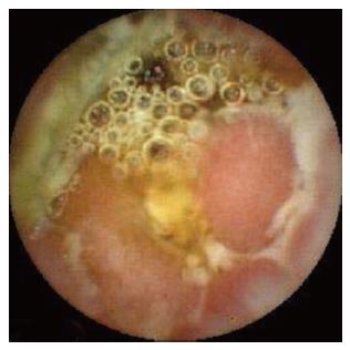
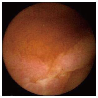
Figure 4 Capsule endoscopy.
Multiple ulcers of the terminal ileum leading to lumen sub-stenosis.
Figure 5 Capsule endoscopy.
Linear ulcer of the jejunum.
- Citation: Saibeni S, Rondonotti E, Iozzelli A, Spina L, Tontini GE, Cavallaro F, Ciscato C, de Franchis R, Sardanelli F, Vecchi M. Imaging of the small bowel in Crohn's disease: A review of old and new techniques. World J Gastroenterol 2007; 13(24): 3279-3287
- URL: https://www.wjgnet.com/1007-9327/full/v13/i24/3279.htm
- DOI: https://dx.doi.org/10.3748/wjg.v13.i24.3279


