Copyright
©2007 Baishideng Publishing Group Co.
World J Gastroenterol. Jun 7, 2007; 13(21): 2986-2991
Published online Jun 7, 2007. doi: 10.3748/wjg.v13.i21.2986
Published online Jun 7, 2007. doi: 10.3748/wjg.v13.i21.2986
Figure 1 Representative AP-PCR fingerprints of ICC (T) and corresponding normal tissue DNA (N), with primer BC17.
Arrowhead indicates the position of altered DNA fragments.
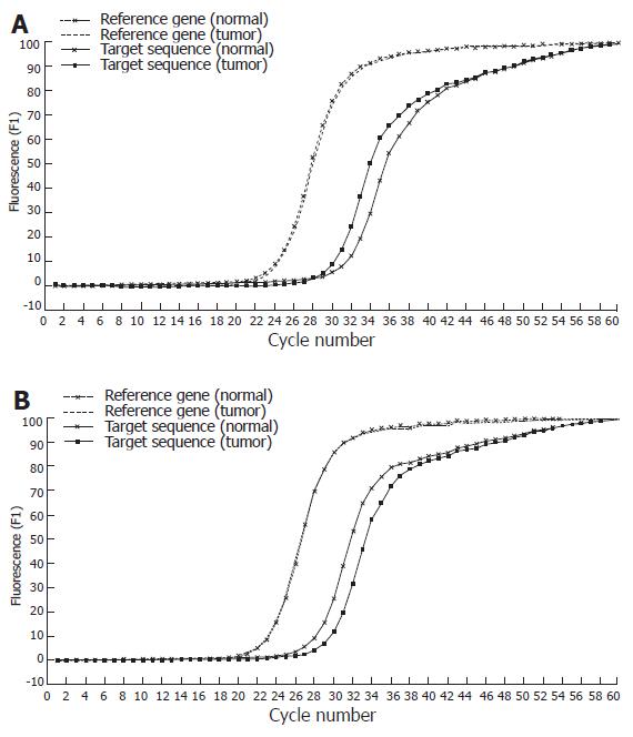
Figure 2 Real-time PCR SYBR Green I fluorescence record versus cycle number of target fragment (518bp) and reference gene (β-globin) in tumor DNA and corresponding normal DNA, in case of DNA amplification (A) and allelic loss (B).
Figure 3 Changes in DNA copy number of ICC (T) and corresponding normal tissue DNA (N) compared with reference gene (β-globin).
Case 11 showed no change. Cases 23 and 31 showed loss of DNA copy number, whereas cases 26 and 45 showed DNA amplification at chromosome 17p13.2.
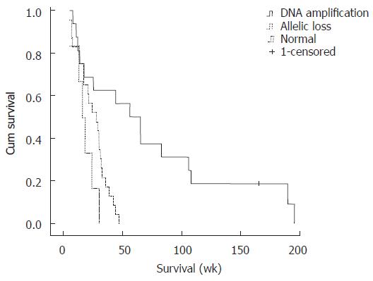
Figure 4 Kaplan-Meier estimated survival rates according to the altered DNA fragment on chromosome 17p13.
2.
- Citation: Chuensumran U, Wongkham S, Pairojkul C, Chauin S, Petmitr S. Prognostic value of DNA alterations on chromosome 17p13.2 for intrahepatic cholangiocarcinoma. World J Gastroenterol 2007; 13(21): 2986-2991
- URL: https://www.wjgnet.com/1007-9327/full/v13/i21/2986.htm
- DOI: https://dx.doi.org/10.3748/wjg.v13.i21.2986


