Copyright
©2007 Baishideng Publishing Group Co.
World J Gastroenterol. Jun 7, 2007; 13(21): 2918-2922
Published online Jun 7, 2007. doi: 10.3748/wjg.v13.i21.2918
Published online Jun 7, 2007. doi: 10.3748/wjg.v13.i21.2918
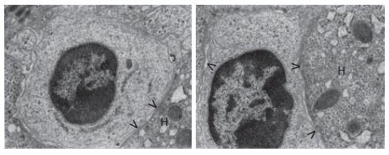
Figure 1 The view of hepatic progenitor cells in hepatic intracellular spaces.
The nuclei contain dense heterochromatin clumped under nuclear envelope and sparse euchromatin. The cytoplasm shows rare poorly developed cell organelles. Between progenitor cells and the neighboring mature hepatic cells (H) point desmosomes are present (>) (× 12 000).
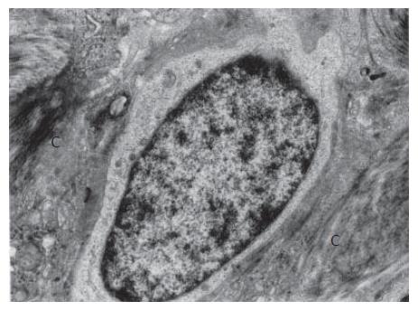
Figure 2 In the field of massive periportal fibrosis, an intermediate hepatocyte-like cell bigger than hepatic progenitor cell, with the electron-lighter cytoplasm and the nucleus with low heterochromatin content and resembling the hepatocyte nucleus.
Cell organelles: a small number and poorly developed. C: collagen fiber bundles (× 7000).
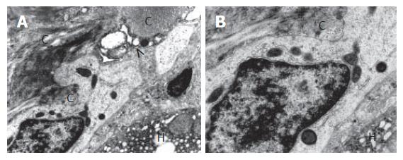
Figure 3 In a fibrotic field of the periportal space; an intermediate hepatic-like cell and transformed Ito cell.
A thick bundle of collagen fibers (C) exerts a pressure on the cell from the outside, causing its focal narrowing; the electron-light cytoplasm contains relatively well developed dark mitochondria and elements of endoplasmic reticulum. Transformed Ito cell (>) adhering to intermediate hepatic-like cell surrounded by collagen deposits (C); H: hepatocyte of the limiting plate of the lobule. A: × 7000; B: × 12 000.
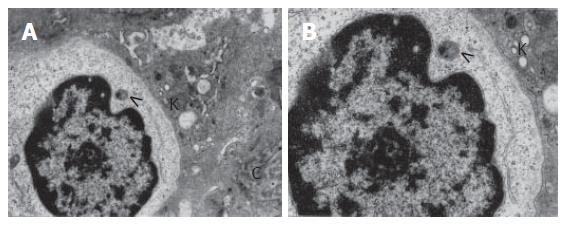
Figure 4 The distended perisinusoidal space of Disse shows an intermediate hepatocyte-like cell with adhering Kupffer cell (K); the IHC nucleus has less nuclear heterochromatin than the HPC nucleus and contains the nucleolus; the low electron dense cytoplasm with a distinct structure that resembles a peroxysome currently being formed (>) and with granular endoplasmic reticulum profiles.
C: a bundle of collagen fibers. A: × 7000; B: × 12 000.
- Citation: Sobaniec-Lotowska ME, Lotowska JM, Lebensztejn DM. Ultrastructure of oval cells in children with chronic hepatitis B, with special emphasis on the stage of liver fibrosis: The first pediatric study. World J Gastroenterol 2007; 13(21): 2918-2922
- URL: https://www.wjgnet.com/1007-9327/full/v13/i21/2918.htm
- DOI: https://dx.doi.org/10.3748/wjg.v13.i21.2918


