Copyright
©2007 Baishideng Publishing Group Co.
World J Gastroenterol. May 28, 2007; 13(20): 2798-2802
Published online May 28, 2007. doi: 10.3748/wjg.v13.i20.2798
Published online May 28, 2007. doi: 10.3748/wjg.v13.i20.2798
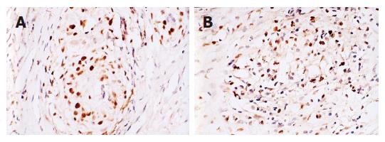
Figure 1 A: The mutated p53 positive expression in tumor tissue (× 200); B: c-myc positive expression in tumor tissue (× 200).
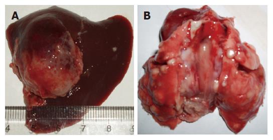
Figure 2 A: The outward appearance of VX2 liver tumor the metastatic carcinoma on lung in control group; B: The metastatic carcinoma on lung in control group.
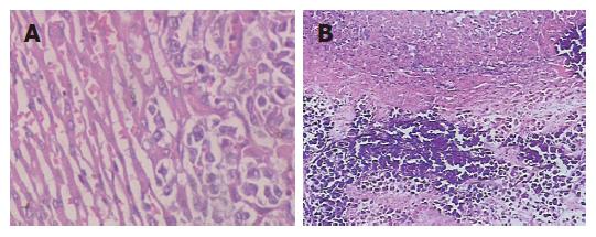
Figure 3 A: There are apparent morphological differences between VX2 liver tumor tissue and the normal liver tissue (× 200); B: There are lots of calcified points in tumor tissue in group treated by nano-HAP (× 100).
- Citation: Hu J, Liu ZS, Tang SL, He YM. Effect of hydroxyapatite nanoparticles on the growth and p53/c-Myc protein expression of implanted hepatic VX2 tumor in rabbits by intravenous injection. World J Gastroenterol 2007; 13(20): 2798-2802
- URL: https://www.wjgnet.com/1007-9327/full/v13/i20/2798.htm
- DOI: https://dx.doi.org/10.3748/wjg.v13.i20.2798


