Copyright
©2007 Baishideng Publishing Group Co.
World J Gastroenterol. Jan 14, 2007; 13(2): 320-323
Published online Jan 14, 2007. doi: 10.3748/wjg.v13.i2.320
Published online Jan 14, 2007. doi: 10.3748/wjg.v13.i2.320
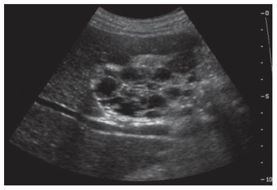
Figure 1 Abdominal ultrasonography showing multi-loculated cystic mass and intact gallbladder lumen.
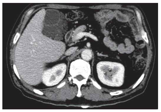
Figure 2 Abdominal computed tomography (CT) image showing a low-density multilocular cystic lesion with septation originated from the gallbladder fossa.
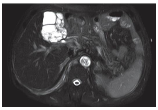
Figure 3 Axial T2-weighted magnetic resonance (MR) image showing a thin multi-septated cystic mass (high-signal intensity) with scalloping margin.
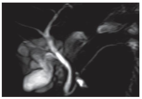
Figure 4 Magnetic resonance cholangiopancreatography (MRCP) image showing gallbladder encapsulated by variable sized multi-cystic lesions, which have no communication with biliary system.
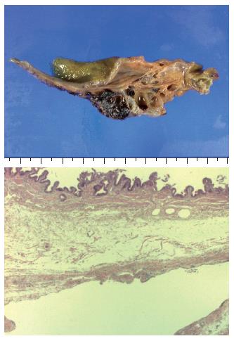
Figure 5 Gross appearance of resected specimen showing a well-marginated multi cystic lesion filled with yellowish fluid, and microphotograph from the cholecystectomy specimen showing a lymphatic space lined with flat endothelium (hematoxylin and eosin staining, x12.
5).
- Citation: Kim JK, Yoo KS, Moon JH, Park KH, Chung YW, Kim KO, Park CH, Hahn T, Park SH, Kim JH, Jeon JY, Kim MJ, Min KS, Park CK. Gallbladder lymphangioma: A case report and review of the literature. World J Gastroenterol 2007; 13(2): 320-323
- URL: https://www.wjgnet.com/1007-9327/full/v13/i2/320.htm
- DOI: https://dx.doi.org/10.3748/wjg.v13.i2.320


