Copyright
©2007 Baishideng Publishing Group Co.
World J Gastroenterol. Jan 14, 2007; 13(2): 316-319
Published online Jan 14, 2007. doi: 10.3748/wjg.v13.i2.316
Published online Jan 14, 2007. doi: 10.3748/wjg.v13.i2.316
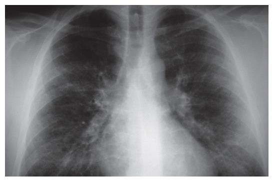
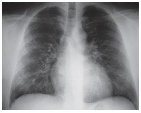
Figure 1 Chest X-ray (2003-09-09) showing moderate reticular enhancement with ring-like consolidation in both lungs (but predominantly the right), without cardiac or aortic abnormalities.
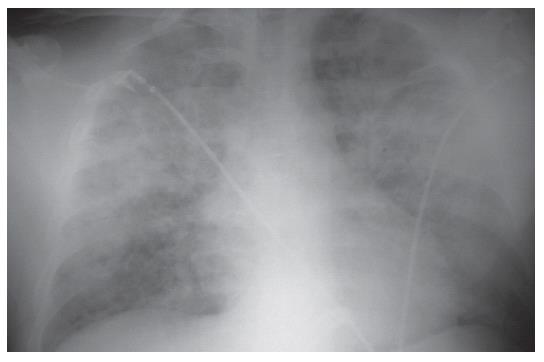
Figure 2 Chest X-ray (2003-09-14) showing significant progression and volume loss in both lungs.
A palm-sized homogeneous consolidation developed in the central part of the lung, a marked interstitial enhancement was seen in other parts of the lung. The radiological image suggested ARDS. The heart was enlarged.
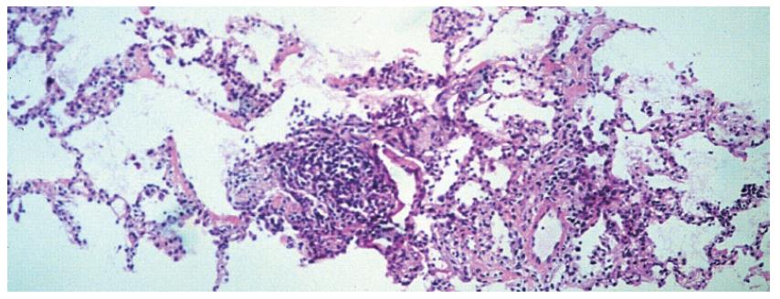
Figure 3 Histology of lung needle biopsy (HE, 112 ×).
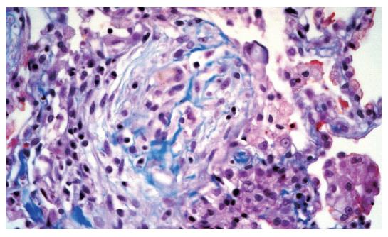
Figure 4 Histology showing the pathognomic Masson bodies for BOOP and polypous proliferation of new connective tissue in the alveolus (trichrome, 224 ×).
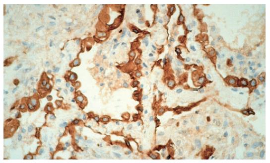
Figure 5 Immunohistochemistry for the expression of CK 7.
Interstitial fibrosis and proliferation of type II pneumocytes are apparent (224 ×).
Figure 6 Chest X-ray (2003-10-15) showing improvement of lung volumes.
Reticular enhancement of the middle part of the lungs is still present.
- Citation: Nagy F, Molnar T, Makula E, Kiss I, Milassin P, Zollei E, Tiszlavicz L, Lonovics J. A case of interstitial pneumonitis in a patient with ulcerative colitis treated with azathioprine. World J Gastroenterol 2007; 13(2): 316-319
- URL: https://www.wjgnet.com/1007-9327/full/v13/i2/316.htm
- DOI: https://dx.doi.org/10.3748/wjg.v13.i2.316


