Copyright
©2007 Baishideng Publishing Group Co.
World J Gastroenterol. Jan 14, 2007; 13(2): 310-312
Published online Jan 14, 2007. doi: 10.3748/wjg.v13.i2.310
Published online Jan 14, 2007. doi: 10.3748/wjg.v13.i2.310
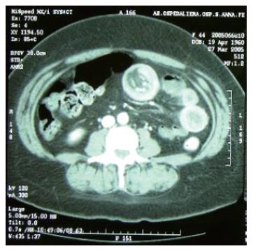
Figure 1 Duodenum-jejunal distension and the presence of a jejunal loop with thickened walls, with a second hyper-dense image inside the same lumen, forming a target-shaped aspect characteristic of intestinal invagination.
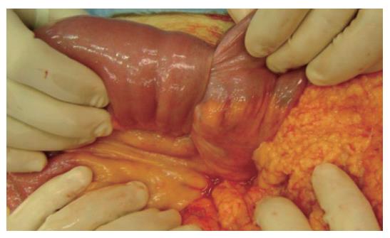
Figure 2 Invagination at the third jejunal loop.
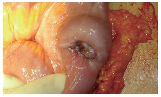
Figure 3 Presence of a hyperchromic ulcerated neoformation, with signs of recent bleeding of the serosa.
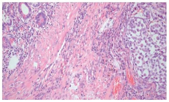
Figure 4 Ileal wall infiltrated with metastatic melanomatous cells with nuclear pseudoinclusions and nucleoli, in nest and trabecular arrangement (HE x 10).
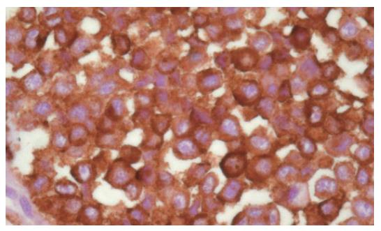
Figure 5 Strong positivity at immunohistochemical assay for HMB45.
- Citation: Resta G, Anania G, Messina F, de Tullio D, Ferrocci G, Zanzi F, Pellegrini D, Stano R, Cavallesco G, Azzena G, Occhionorelli S. Jejuno-jejunal invagination due to intestinal melanoma. World J Gastroenterol 2007; 13(2): 310-312
- URL: https://www.wjgnet.com/1007-9327/full/v13/i2/310.htm
- DOI: https://dx.doi.org/10.3748/wjg.v13.i2.310


