Copyright
©2007 Baishideng Publishing Group Co.
World J Gastroenterol. Apr 21, 2007; 13(15): 2247-2249
Published online Apr 21, 2007. doi: 10.3748/wjg.v13.i15.2247
Published online Apr 21, 2007. doi: 10.3748/wjg.v13.i15.2247
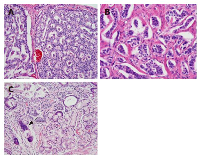
Figure 1 Histological examination of each carcinoid in three organs.
A: Duodenal carcinoid showing anastomotic ribbon-like formation in the interstitium with predominant blood vessels and only a small amount of connective tissue (HE, × 10); B: A palisade-like carcinoid in the pancreas, and a moderate amount of fibrous connective tissue observed in the interstitium (HE, × 20); C: Vesicular carcinoids in the stomach (arrow) (HE, × 10).
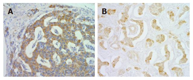
Figure 2 Imunohistochemical staining of carcinoids in the duodenum and pancreas (× 20).
A: Gatrin immunostaining showing a quite positive carcinoid in the duodenum; B: Serotonin immunostaining showing a strongly positive carcinoid in the pancreas.
- Citation: Bamba T, Kosugi SI, Kanda T, Tsubono T, Sakai Y, Musha N, Ishihara N, Hatakeyama K. Multiple carcinoids in the duodenum, pancreas and stomach accompanied with type A gastritis: A case report. World J Gastroenterol 2007; 13(15): 2247-2249
- URL: https://www.wjgnet.com/1007-9327/full/v13/i15/2247.htm
- DOI: https://dx.doi.org/10.3748/wjg.v13.i15.2247


