Copyright
©2007 Baishideng Publishing Group Co.
World J Gastroenterol. Apr 14, 2007; 13(14): 2129-2131
Published online Apr 14, 2007. doi: 10.3748/wjg.v13.i14.2129
Published online Apr 14, 2007. doi: 10.3748/wjg.v13.i14.2129
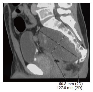
Figure 1 Abdominal CT scan shows a disomogeneous mass arising from the sacral canal through the third right sacral foramen.
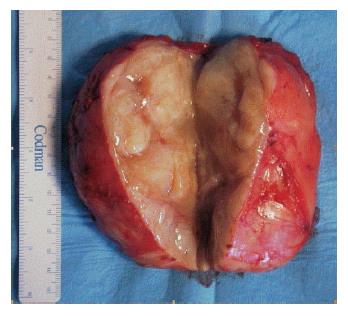
Figure 2 Tumor mass exposed after longitudinal section.
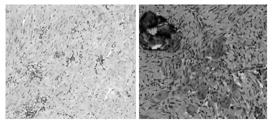
Figure 3 Hematoxylin and Eosin photomicrograph (× 10; × 20).
Large ganglion cells are embedded in a neuromatous stroma with calcifications.
- Citation: Cerullo G, Marrelli D, Rampone B, Miracco C, Caruso S, Marianna DM, Mazzei MA, Roviello F. Presacral ganglioneuroma: A case report and review of literature. World J Gastroenterol 2007; 13(14): 2129-2131
- URL: https://www.wjgnet.com/1007-9327/full/v13/i14/2129.htm
- DOI: https://dx.doi.org/10.3748/wjg.v13.i14.2129


