Copyright
©2007 Baishideng Publishing Group Co.
World J Gastroenterol. Apr 14, 2007; 13(14): 2048-2052
Published online Apr 14, 2007. doi: 10.3748/wjg.v13.i14.2048
Published online Apr 14, 2007. doi: 10.3748/wjg.v13.i14.2048
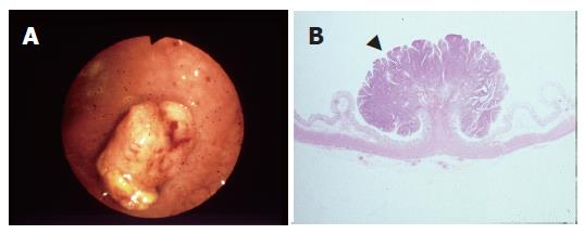
Figure 1 A: Macroscopic appearance of PG submucoal invasive early colorectal carcinoma, note the polyp-like exophytic from of the lesion; B: Microscopic appearance of PG submucoal invasive early colorectal carcinoma, note the intramucosal protruding proliferation (arrow head).
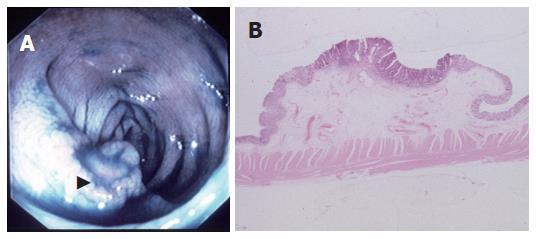
Figure 2 A: Macroscopic appearance of NPG and PG submucoal invasive early colorectal carcinoma, note the depressed from of the lesion (arrow head); B: Microscopic appearance of PG submucoal invasive early colorectal carcinoma, no intramucosal protruding proliferation in the lesion.
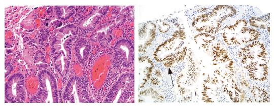
Figure 3 p53 immunohistochemical staining in NPG submucosal invasive early colorectal carcinomas (× 200).
Note positive staining of p53 in cancer tissue (arrows), the picture on the left side is the same section of HE staining.
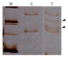
Figure 4 SSCP analysis of the K-ras gene codon 12 in PG submucosal invasive early colorectal carcinomas, note two extra bands of point mutations (arrow head).
C: normal control; T: polypoid submucosal colon cancer; M: molecular marker.
- Citation: Hirata I, Wang FY, Murano M, Inoue T, Toshina K, Nishikawa T, Maemura K. Histopathological and genetic differences between polypoid and non-polypoid submucosal colorectal carcinoma. World J Gastroenterol 2007; 13(14): 2048-2052
- URL: https://www.wjgnet.com/1007-9327/full/v13/i14/2048.htm
- DOI: https://dx.doi.org/10.3748/wjg.v13.i14.2048


