Copyright
©2007 Baishideng Publishing Group Co.
World J Gastroenterol. Mar 21, 2007; 13(11): 1723-1727
Published online Mar 21, 2007. doi: 10.3748/wjg.v13.i11.1723
Published online Mar 21, 2007. doi: 10.3748/wjg.v13.i11.1723
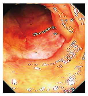
Figure 1 Retrograde ileoscopy showing an ulcer in the terminal ileum.
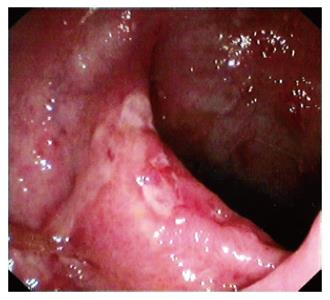
Figure 2 Ulcers and nodularity of the terminal ileal mucosa.
The endoscopic lesions were confined only to the terminal ileum and colonoscopy till the cecum did not reveal any abnormality.
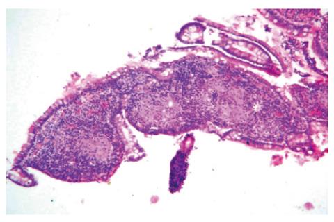
Figure 3 Histological appearance of the mucosal biopsies obtained from the lesion shown in Figure 1.
Note the presence of noncaseating granulomas (HE × 40).
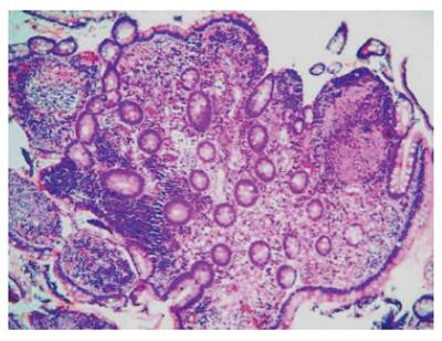
Figure 4 Noncaseating granulomas seen upon histological examination of the mucosal biopsies obtained from the lesion shown in Figure 2 (HE × 80).
- Citation: Misra S, Misra V, Dwivedi M. Ileoscopy in patients with ileocolonic tuberculosis. World J Gastroenterol 2007; 13(11): 1723-1727
- URL: https://www.wjgnet.com/1007-9327/full/v13/i11/1723.htm
- DOI: https://dx.doi.org/10.3748/wjg.v13.i11.1723


