Copyright
©2006 Baishideng Publishing Group Co.
World J Gastroenterol. Mar 7, 2006; 12(9): 1416-1420
Published online Mar 7, 2006. doi: 10.3748/wjg.v12.i9.1416
Published online Mar 7, 2006. doi: 10.3748/wjg.v12.i9.1416
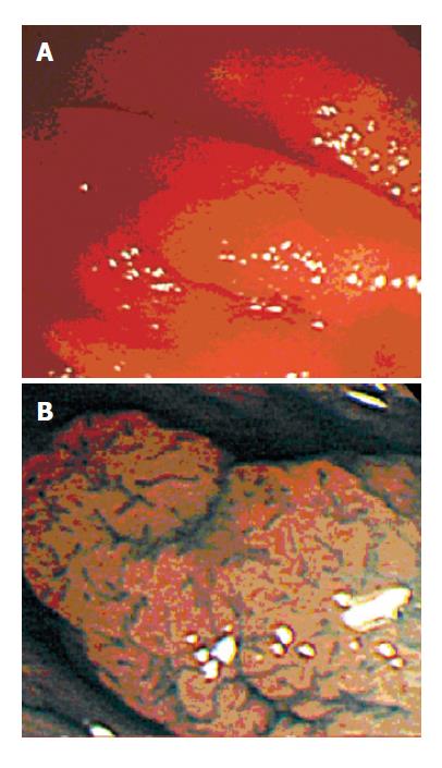
Figure 1 A: Colonoscopy revealed a small reddish polypoid lesion 3 mm in diameter.
B: Chromoendoscopy with magnification disclosed a type II pit, and the lesion was diagnosed as non-neoplastic and examined at biopsy. The histologic diagnosis was hyperplastic polyp.
Figure 2 A: Colonoscopy revealed a small flat lesion 5 mm in diameter.
B: Chromoendoscopy with magnification disclosed a type IIIL pit, and the lesion was diagnosed as neoplastic and resected by snare polypectomy. The histologic diagnosis was adenoma with moderate atypia.
- Citation: Kato S, Fu KI, Sano Y, Fujii T, Saito Y, Matsuda T, Koba I, Yoshida S, Fujimori T. Magnifying colonoscopy as a non-biopsy technique for differential diagnosis of non-neoplastic and neoplastic lesions. World J Gastroenterol 2006; 12(9): 1416-1420
- URL: https://www.wjgnet.com/1007-9327/full/v12/i9/1416.htm
- DOI: https://dx.doi.org/10.3748/wjg.v12.i9.1416


