Copyright
©2006 Baishideng Publishing Group Co.
World J Gastroenterol. Feb 28, 2006; 12(8): 1211-1218
Published online Feb 28, 2006. doi: 10.3748/wjg.v12.i8.1211
Published online Feb 28, 2006. doi: 10.3748/wjg.v12.i8.1211
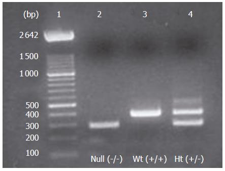
Figure 1 Genotyping of animal offsprings by PCR of cDNA (tail extracts).
Lane 1= molecular weight ladder of 100 bp; lane 2= null mice; lane 3= wild-type (Wt) mice; lane 4 = heterozygous(Ht) mice.
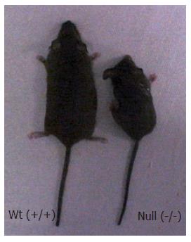
Figure 2 Morphology of Smad3 null and wild-type mice.
The majority of Smad3 null mice exhibited a reduced size compared to littermate controls. Severe bending of forepaw joints was present in Smad3 null mice.
Figure 3 Body Weight changes of wild-type and Smad3 null mice.
Each point represents mean weight data pooled from 10 mice. Standard deviations are indicated. Plot of weight (g) vs age (days). Wild-type are indicated as □ (squares), and null as ◊ (diamonds).
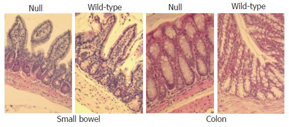
Figure 4 Haematoxylin and eosin-stained sections (x 20) analysis of small and large bowel from wild-type and Smad3 null mice shows normal morphology.
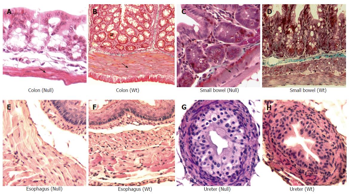
Figure 5 Masson trichrome staining (x 20) of small and large bowel from Smad3 mice.
Significant reduction of muscular layer of descending colon of Smad3 null (A) is observed compared to colon from wild-type (WT) mice (B), reduction of muscle layer in cross sections of the proximal small bowel from Smad3 null (C) as compared to wild-type mice (D). Haematoxylin and eosin staining (x 20) of ureter and esophagus of Smad3 mice. Cross section of esophagus from Smad3 null (E) and wild-type mice (F) shows no differences in muscle layer. Cross section of ureter from Smad3 null (G) and wild-type mice (H) shows no differences in muscle layers.
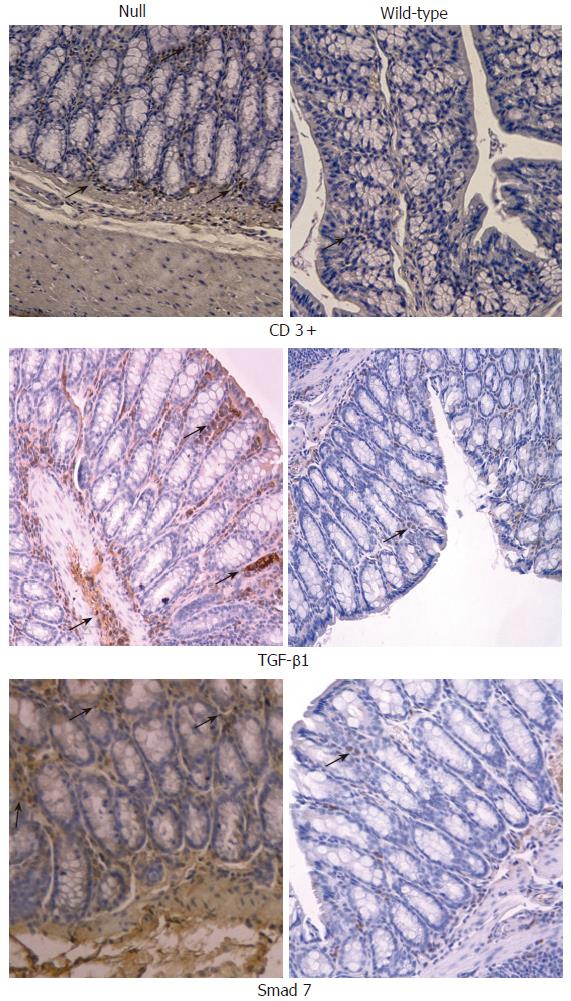
Figure 6 Immunohistochemical analysis (x 20) of CD3+ T cells, TGF- β1 and Smad7 in colon obtained from Smad3 null and wild-type mice.
CD3+ T cells were significantly increased within large intestine of Smad3 null mice as compared to the wild-type mice. TGF-β1 and Smad7 were significantly increased within large intestine of Smad3 null mice compared to wild-type mice.
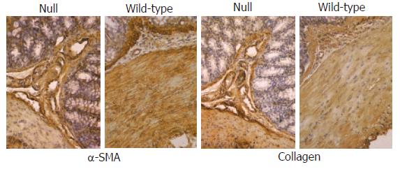
Figure 7 Immunohistochemical analysis (x 20) of α-SMA and collagens I-VII in colon from Smad3 null and wild-type mice.
A similar localization of α-SMA antibody was found in miocytes of muscularis mucosae, muscle layer and vessels of both groups of animals. Staining of collagens I-VII in large intestine of Smad3 null and wild-type mice was localized mainly within connective tissue of submucosa and muscularis propria showing identical staining pattern between the two groups of mice.
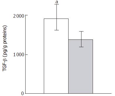
Figure 8 TGF- β1 ELISA of colon homogenates from Smad3 null (solid column) and wild-type mice (dashed column).
Data are given as mean±SD. aP < 0.05 vs wild type mice.
- Citation: Zanninelli G, Vetuschi A, Sferra R, D’Angelo A, Fratticci A, Continenza MA, Chiaramonte M, Gaudio E, Caprilli R, Latella G. Smad3 knock-out mice as a useful model to study intestinal fibrogenesis. World J Gastroenterol 2006; 12(8): 1211-1218
- URL: https://www.wjgnet.com/1007-9327/full/v12/i8/1211.htm
- DOI: https://dx.doi.org/10.3748/wjg.v12.i8.1211


