Copyright
©2006 Baishideng Publishing Group Co.
World J Gastroenterol. Feb 14, 2006; 12(6): 977-978
Published online Feb 14, 2006. doi: 10.3748/wjg.v12.i6.977
Published online Feb 14, 2006. doi: 10.3748/wjg.v12.i6.977
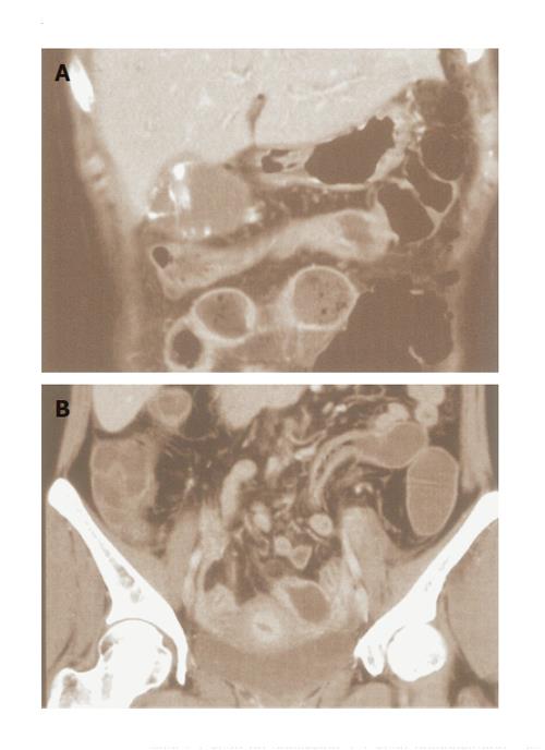
Figure 1 Abdominal CT scan on admission.
CT scan demonstrated gallbladder swelling with thickened wall and stones, ascites, a thickened ileum, and fluid collection in the small intestine.
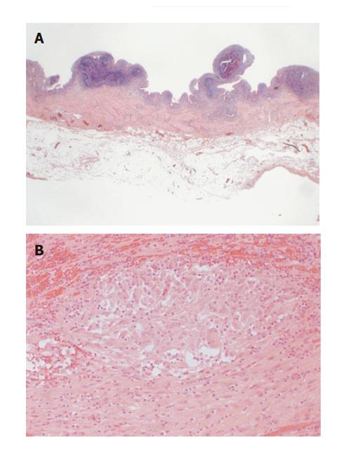
Figure 2 Histological findings of the gallbladder.
A: The mucosa was nodular and granular, and the wall was thickened. Erosion and submucosal bleeding were also observed, but there were no obvious ulcers; B: There were several well-formed epithelioid cell granulomas, which was non-caseating sarcoidal type, different from the foreign-body and xanthomatous granulomas.
- Citation: Andoh A, Endo Y, Kushima R, Hata K, Tsujikawa T, Sasaki M, Mekata E, Tani T, Fujiyama Y. A case of Crohn’s disease involving the gallbladder. World J Gastroenterol 2006; 12(6): 977-978
- URL: https://www.wjgnet.com/1007-9327/full/v12/i6/977.htm
- DOI: https://dx.doi.org/10.3748/wjg.v12.i6.977


