Copyright
©2006 Baishideng Publishing Group Co.
World J Gastroenterol. Feb 7, 2006; 12(5): 800-803
Published online Feb 7, 2006. doi: 10.3748/wjg.v12.i5.800
Published online Feb 7, 2006. doi: 10.3748/wjg.v12.i5.800
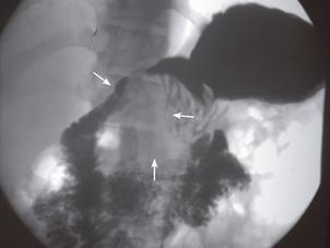
Figure 1 Upper gastrointestinal series shows that the gastric antrum (arrows) is deformed due to external compression.
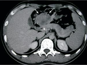
Figure 2 Abdominal computed tomography shows a 5 cm heterogeneous mass (arrows) located between the gastric antrum, duodenal loop, and pancreatic head.
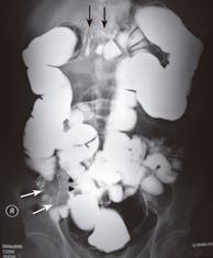
Figure 3 Barium enema study demonstrates a mass compressing the middle transverse colon with tethering appearance, suggesting gastrocolic ligament invasion (black arrows).
Long segmental encasement of cecum, proximal ascending colon (white arrows), and ileocecal valve (arrows head) is also evident.
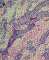
Figure 4 A: Photomicrography depicts nests or clusters of small tumor cells outlined by characteristic desmoplastic stroma bands (hematoxylin and eosin staining, 100×).
B: Photomicrography demonstrates the monomorphic, small round tumor cell population with a small spherical or spheroid nucleus, rich in chromatin (hematoxylin and eosin staining, 400×). C: The immunohistochemical staining of tumor cells is positive for desmin.
- Citation: Chang CC, Hsu JT, Tseng JH, Hwang TL, Chen HM, Jan YY. Combined resection and multi-agent adjuvant chemotherapy for desmoplastic small round cell tumor arising in the abdominal cavity: Report of a case. World J Gastroenterol 2006; 12(5): 800-803
- URL: https://www.wjgnet.com/1007-9327/full/v12/i5/800.htm
- DOI: https://dx.doi.org/10.3748/wjg.v12.i5.800


