Copyright
©2006 Baishideng Publishing Group Co.
World J Gastroenterol. Dec 28, 2006; 12(48): 7874-7877
Published online Dec 28, 2006. doi: 10.3748/wjg.v12.i48.7874
Published online Dec 28, 2006. doi: 10.3748/wjg.v12.i48.7874
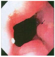
Figure 1 Initial endoscopic image of localized nodularity in esophageal mucosal surface with irregular Z-line.
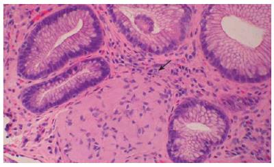
Figure 2 Hematoxylin and eosin stained tissue section of biopsy from irregular EG-Junction showing neuroid proliferation within lamina propria (× 10).
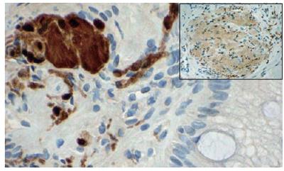
Figure 3 Focal positivity for synaptophysin (insert × 10) confirms the presence of a neuronal (ganglio) component.
S100 (× 20) confirms nerve sheath-neuromatous/neuromatosis component.
- Citation: Siderits R, Hanna I, Baig Z, Godyn JJ. Sporadic ganglioneuromatosis of esophagogastric junction in a patient with gastro-esophageal reflux disorder and intestinal metaplasia. World J Gastroenterol 2006; 12(48): 7874-7877
- URL: https://www.wjgnet.com/1007-9327/full/v12/i48/7874.htm
- DOI: https://dx.doi.org/10.3748/wjg.v12.i48.7874


