Copyright
©2006 Baishideng Publishing Group Co.
World J Gastroenterol. Dec 21, 2006; 12(47): 7654-7659
Published online Dec 21, 2006. doi: 10.3748/wjg.v12.i47.7654
Published online Dec 21, 2006. doi: 10.3748/wjg.v12.i47.7654
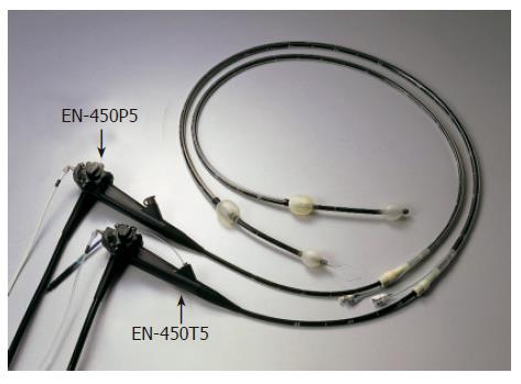
Figure 1 Two types of double-balloon videoenteroscopes (EN-450P5 and EN-450T5).
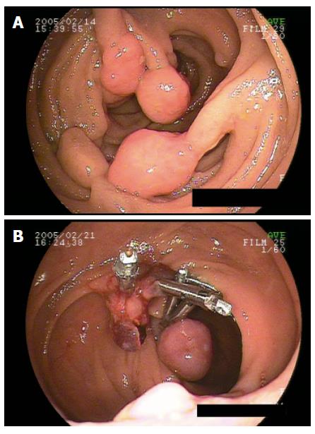
Figure 2 The successful endoscopic removal of polyps from the mid small bowel in a patient with Peutz-Jeghers syndrome.
A: Endoscopic view of multiple pedunculated small intestinal polyps; B: Endoscopic view of the region after endoscopic polypectomy using clipping.
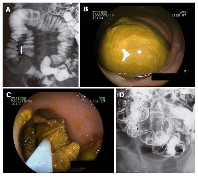
Figure 3 Successful endoscopic lithotripsy for a huge enterolithiasis.
A: Radiographic view of the ileum showing a huge enterolith (arrow); B: Endoscopic view of a huge enterolith; C: Endoscopic view of the lesion during lithotripsy; D: Radiographic view of the region after endoscopic lithotripsy showing no enterolith (arrow).
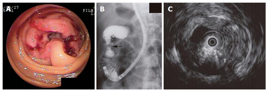
Figure 4 Primary advanced jejunal cancer.
A: Endoscopic image; B: Selective radiographic image using DBE revealing a stenotic lesion (arrow) suggestive of jejunal cancer; C: EUS image using ultrasound catheter probe showing a hypoechoic tumor (T), which extended into the serosal layer (T3).
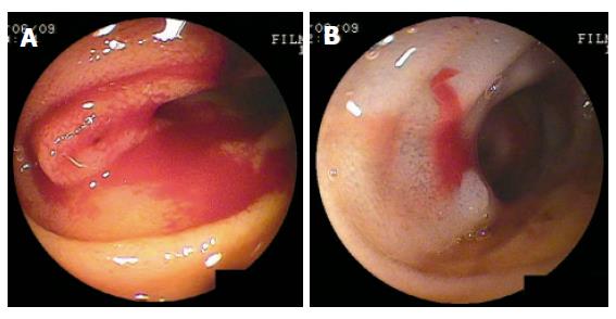
Figure 5 Endoscopic hemostasis using hypertonic-saline solution epinephrine injection.
A: Endoscopic image of bleeding angiodysplasia; B: Endoscopic image of the region after hemostasis.
- Citation: Akahoshi K, Kubokawa M, Matsumoto M, Endo S, Motomura Y, Ouchi J, Kimura M, Murata A, Murayama M. Double-balloon endoscopy in the diagnosis and management of GI tract diseases: Methodology, indications, safety, and clinical impact. World J Gastroenterol 2006; 12(47): 7654-7659
- URL: https://www.wjgnet.com/1007-9327/full/v12/i47/7654.htm
- DOI: https://dx.doi.org/10.3748/wjg.v12.i47.7654


