Copyright
©2006 Baishideng Publishing Group Co.
World J Gastroenterol. Nov 28, 2006; 12(44): 7210-7212
Published online Nov 28, 2006. doi: 10.3748/wjg.v12.i44.7210
Published online Nov 28, 2006. doi: 10.3748/wjg.v12.i44.7210
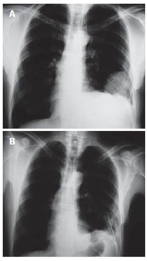
Figure 1 (A) Preopera-tive chest radiograph showing the left-sided opacity and (B) postoperative chest radiograph showing the chest tube placed in the left thoracic cavity.
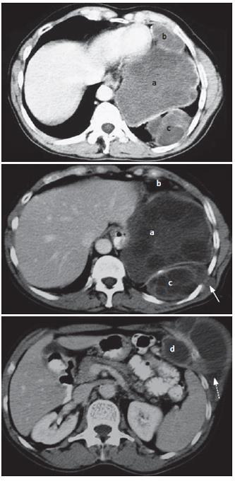
Figure 2 Thoraco-abdominal CT scan showing four separate hydatid cysts located in the left hemidiaphragm (a), anteriorly in contact with the left side of the pericardium (b) and the posterolateral wall of the left hemithorax (c) and intra-abdominally in contact with the left hypochondrium (d).
Subcutaneous extension of hydatid cyst c protruding intercostally to a small subcutaneous cyst under the scapulae (solid arrow) and abdominal cyst d protruding through the left costal cartilage to the left hypochondrium subcutaneously (broken arrow) are demonstrated.
- Citation: Marinis A, Fragulidis G, Karapanos K, Konstantinidis C, Brestas P, Vassiliou J, Smyrniotis V. Subcutaneous extension of a large diaphragmatic hydatid cyst. World J Gastroenterol 2006; 12(44): 7210-7212
- URL: https://www.wjgnet.com/1007-9327/full/v12/i44/7210.htm
- DOI: https://dx.doi.org/10.3748/wjg.v12.i44.7210


