Copyright
©2006 Baishideng Publishing Group Co.
World J Gastroenterol. Nov 21, 2006; 12(43): 7061-7063
Published online Nov 21, 2006. doi: 10.3748/wjg.v12.i43.7061
Published online Nov 21, 2006. doi: 10.3748/wjg.v12.i43.7061
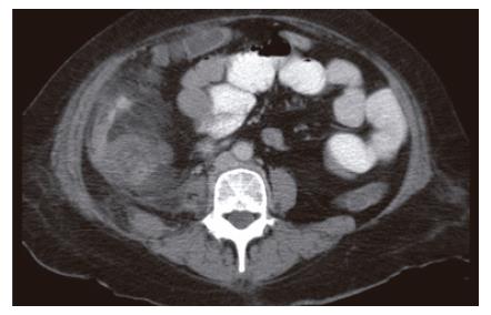
Figure 1 CT of Abdomen showing inflammation around right colon and extravasation of oral contrast.
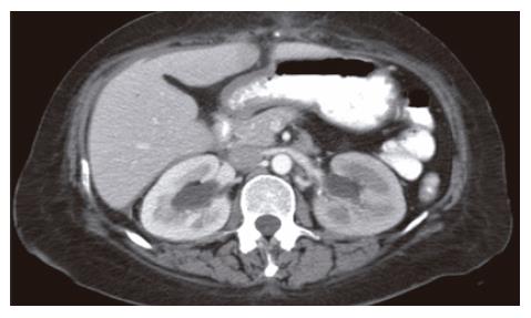
Figure 2 CT of abdomen showing bilateral hydronephrosis.
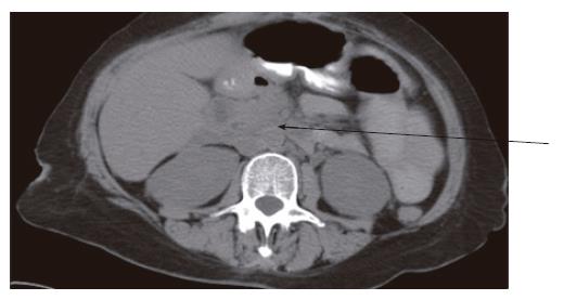
Figure 3 CT of abdomen showing retroperitoneal fibrosis, encasing aorta and inferior vena cava.
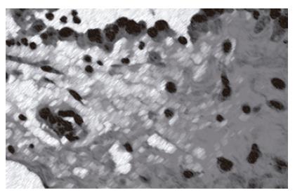
Figure 4 Hematoxylin and Eosin Staining showing fibrinous adhesions and inflammatory reaction.
- Citation: Aziz F, Conjeevaram S, Phan T. Retroperitoneal fibrosis: A rare cause of both ureteral and small bowel obstruction. World J Gastroenterol 2006; 12(43): 7061-7063
- URL: https://www.wjgnet.com/1007-9327/full/v12/i43/7061.htm
- DOI: https://dx.doi.org/10.3748/wjg.v12.i43.7061


