Copyright
©2006 Baishideng Publishing Group Co.
World J Gastroenterol. Nov 21, 2006; 12(43): 7058-7060
Published online Nov 21, 2006. doi: 10.3748/wjg.v12.i43.7058
Published online Nov 21, 2006. doi: 10.3748/wjg.v12.i43.7058
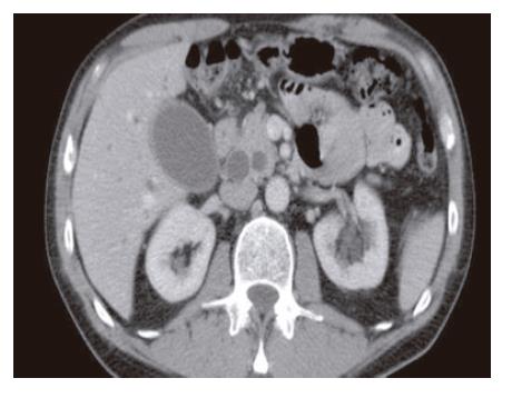
Figure 1 The characteristic “double duct sign” on abdominal CT is indicative of obstruction at the level of the ampulla.
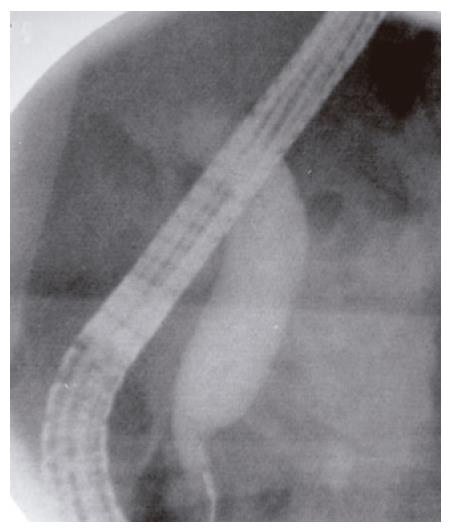
Figure 2 ERCP showing a markedly dilated CBD with an abrupt “shoulder” in the region of the ampulla, suggestive of a periampullary mass.
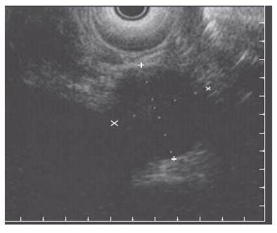
Figure 3 EUS identifying a 23 mm x 27 mm well circumscribed, round, hypoechoic mass in the region of the ampulla, which is distinct from the duodenal wall.
- Citation: Poultsides GA, Frederick WA. Carcinoid of the ampulla of Vater: Morphologic features and clinical implications. World J Gastroenterol 2006; 12(43): 7058-7060
- URL: https://www.wjgnet.com/1007-9327/full/v12/i43/7058.htm
- DOI: https://dx.doi.org/10.3748/wjg.v12.i43.7058


