Copyright
©2006 Baishideng Publishing Group Co.
World J Gastroenterol. Nov 14, 2006; 12(42): 6874-6878
Published online Nov 14, 2006. doi: 10.3748/wjg.v12.i42.6874
Published online Nov 14, 2006. doi: 10.3748/wjg.v12.i42.6874
Figure 1 Photomicrographs of immunoreactive endocrine cells in the gastrointestinal tract of the flower fish.
A: Bombesin immunoreactive cells in the enlarged area after oesophagus; B: Somatostatin immunoreactive cells in the enlarged area after oesophagus; C: Substance-P immunoreactive cells in the enlarged area after oesophagus; D: Vasoactive intestinal peptide immunoreactive cells in the anterior intestine (× 450).
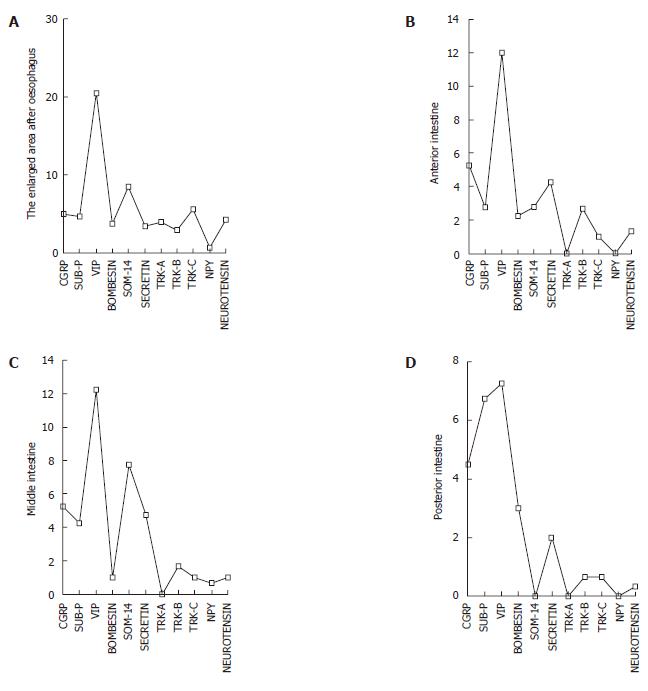
Figure 2 Density of the immunoreactive endocrine cells in the enlarged area after oesophagus (A), the anterior intestine (B), the middle intestine (C) and the posterior intestine (D).
- Citation: Çınar K, Şenol N, Özen MR. Immunohistochemical study on distribution of endocrine cells in gastrointestinal tract of flower fish (Pseudophoxinus antalyae). World J Gastroenterol 2006; 12(42): 6874-6878
- URL: https://www.wjgnet.com/1007-9327/full/v12/i42/6874.htm
- DOI: https://dx.doi.org/10.3748/wjg.v12.i42.6874


