Copyright
©2006 Baishideng Publishing Group Co.
World J Gastroenterol. Oct 28, 2006; 12(40): 6561-6563
Published online Oct 28, 2006. doi: 10.3748/wjg.v12.i40.6561
Published online Oct 28, 2006. doi: 10.3748/wjg.v12.i40.6561
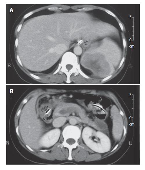
Figure 1 Contrast-enhanced computed tomography of the abdomen.
A: Enlarged spleen with hypodense lesion in the superior spleen which was considered to be splenic metastasis; B: Hypoattenuating thrombus in the splenic vein and the portal vein.
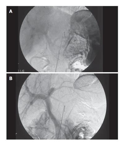
Figure 2 Digital substraction angiography.
A: The venous phase of the celiac artery angiogram reveals occlusion of the splenic vein and development of collaterals; B: The venous phase of the superior mesenteric artery angiogram reveals a defect shadow in the portal vein at its junction with the splenic vein.
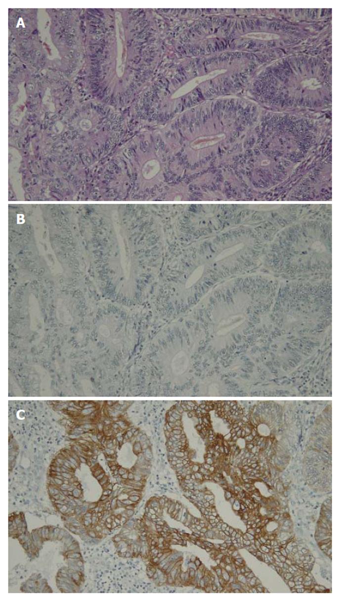
Figure 3 Histological findings of the tumor of the ascending colon.
A: Moderately differentiated adenocarcinoma (HE, x 20); B: Immunostaining for cytokeratin 7 showing a negative reaction (x 20); C: Immunostaining for cytokeratin 20 showing a positive reaction (x 20).
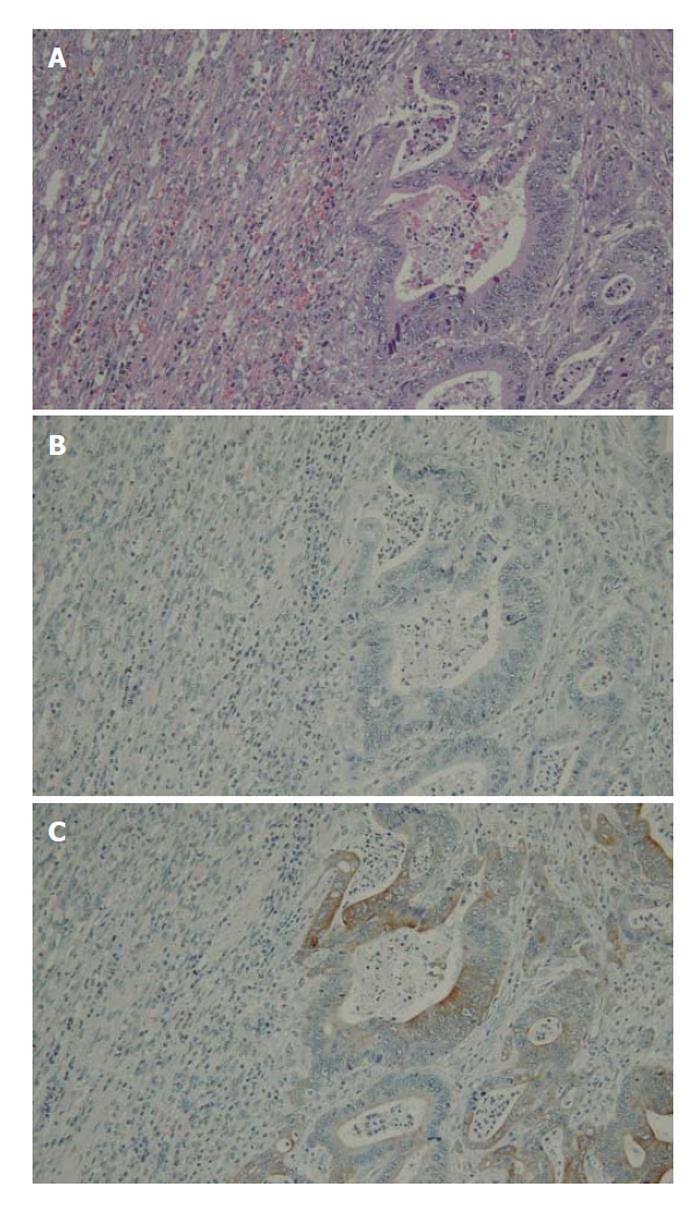
Figure 4 Histological findings of the tumor of the spleen.
A: Moderately differentiated adenocarcinoma (HE x 20); B: Immunostaining for cytokeratin 7 showing a negative reaction (x 20); C: Immunostaining for cytokeratin 20 showing a positive reaction (x 20).
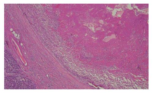
Figure 5 Histological findings of the splenic vein filled with not tumor thrombus, but organizing thrombus.
The elastic tissue ring is intact (HE, x 4).
- Citation: Hiraiwa K, Morozumi K, Miyazaki H, Sotome K, Furukawa A, Nakamaru M, Tanaka Y, Iri H. Isolated splenic vein thrombosis secondary to splenic metastasis: A case report. World J Gastroenterol 2006; 12(40): 6561-6563
- URL: https://www.wjgnet.com/1007-9327/full/v12/i40/6561.htm
- DOI: https://dx.doi.org/10.3748/wjg.v12.i40.6561


