Copyright
©2006 Baishideng Publishing Group Co.
World J Gastroenterol. Oct 28, 2006; 12(40): 6507-6514
Published online Oct 28, 2006. doi: 10.3748/wjg.v12.i40.6507
Published online Oct 28, 2006. doi: 10.3748/wjg.v12.i40.6507
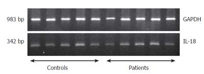
Figure 1 Analysis of RT-PCR amplification of IL-18 in PBMC of CP patients.
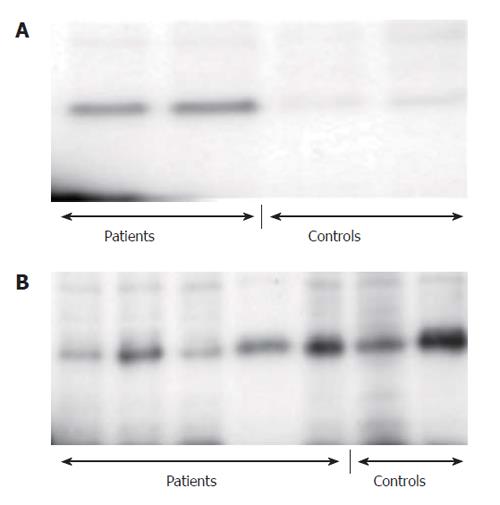
Figure 2 IL-18 in cell lysates of PBMC and in pancreatic tissue of CP patients.
A: In the pancreatic tissue; B: In cell lysates of PBMC.
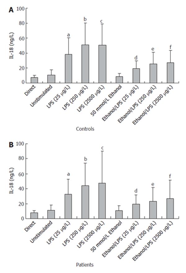
Figure 3 In vitro IL-18 secretion from PBMC of CP patients.
aP < 0.0007 25 μg/L LPS vs unstimulated; bP < 0.0007 250 μg/L LPS vs unstimulated; cP < 0.0007 2500 μg/L LPS vs unstimulated; dP < 0.0001 Ethanol/25 μg/L LPS vs 25 μg/L LPS; eP < 0.0001 Ethanol/250 μg/L LPS vs 250 μg/L LPS; fP < 0.0001 Ethanol/2500 μg/L LPS vs 2500 μg/L LPS.
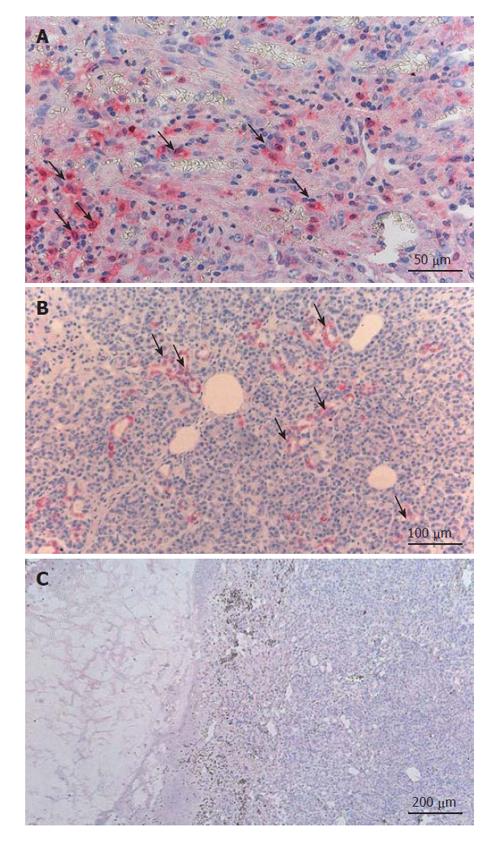
Figure 4 IL-18 expression in pancreatic tissue of CP patients.
A: Clusters of infiltrating mononuclear cells stained positive; B: Clusters of pancreatic acinar cells also stained positive for the expression of IL-18; C: No positive staining for IL-18 in the negative control (mouse serum with IL-18 antigen-antibody mixture).
- Citation: Schneider A, Haas SL, Hildenbrand R, Siegmund S, Reinhard I, Nakovics H, Singer MV, Feick P. Enhanced expression of interleukin-18 in serum and pancreas of patients with chronic pancreatitis. World J Gastroenterol 2006; 12(40): 6507-6514
- URL: https://www.wjgnet.com/1007-9327/full/v12/i40/6507.htm
- DOI: https://dx.doi.org/10.3748/wjg.v12.i40.6507


