Copyright
©2006 Baishideng Publishing Group Co.
World J Gastroenterol. Oct 21, 2006; 12(39): 6397-6400
Published online Oct 21, 2006. doi: 10.3748/wjg.v12.i39.6397
Published online Oct 21, 2006. doi: 10.3748/wjg.v12.i39.6397
Figure 1 Dynamic computed tomography scan showing swelling of the gallbladder and slight swelling of the pancreas head (white arrow) (A).
The pancreas body and tail are not swollen (B). A tumor is detected in the right kidney (C).
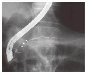
Figure 2 Endoscopic retrograde cholangiopancreatography showing short irregular narrowing of the main pancreatic duct in the head.
The narrowing is less than one third of the length of the main pancreatic duct (white arrowheads).
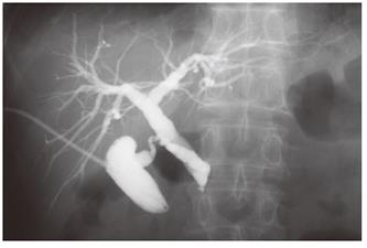
Figure 3 Percutaneous transhepatic gallbladder drainage.
Fluorography through the biliary drainage catheter shows obstruction of the distal common bile duct.
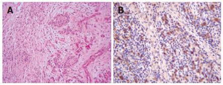
Figure 4 Wedge biopsy of the pancreas head reveals lymphoplasmacytic infiltration and interstitial fibrosis with acinar atrophy (HE x 100) (A).
Immunohistologic study shows dense IgG4-positive plasma cell infiltration in peripancreatic lymph nodes (HE x 200) (B).
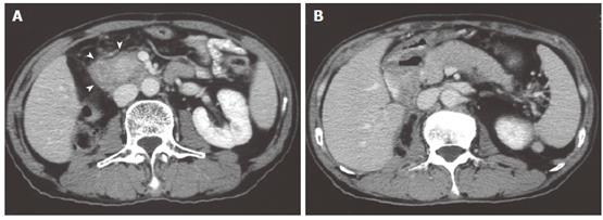
Figure 5 Dynamic computed tomography scan showing swelling of the pancreas head.
A capsule-like low-density rim can be seen around the pancreas head (white arrowheads) (A). The pancreas body and tail are swollen (B).
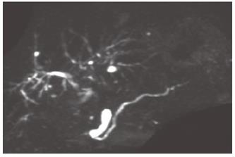
Figure 6 Magnetic resonance cholangiopancreatography performed 4 years after steroid therapy.
The narrowing site is resolved in the main pancreatic duct.
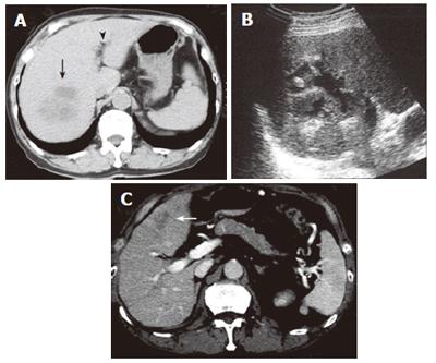
Figure 7 Computed tomography scan showing an abscess in the right anteroinferior segment of the liver (black arrow).
Pneumobilia is detected (black arrowhead) (A). Ultrasonography shows partial liquefaction in the abscess (B). Computed tomography scan showing an abscess in the left medial segment of the liver (white arrow) (C). Note that the pancreas is not swollen.
- Citation: Toshikuni N, Kai K, Sato S, Kitano M, Fujisawa M, Okushin H, Morii K, Takagi S, Takatani M, Morishita H, Uesaka K, Yuasa S. Pyogenic liver abscess after choledochoduodenostomy for biliary obstruction caused by autoimmune pancreatitis. World J Gastroenterol 2006; 12(39): 6397-6400
- URL: https://www.wjgnet.com/1007-9327/full/v12/i39/6397.htm
- DOI: https://dx.doi.org/10.3748/wjg.v12.i39.6397


