Copyright
©2006 Baishideng Publishing Group Co.
World J Gastroenterol. Oct 14, 2006; 12(38): 6219-6224
Published online Oct 14, 2006. doi: 10.3748/wjg.v12.i38.6219
Published online Oct 14, 2006. doi: 10.3748/wjg.v12.i38.6219
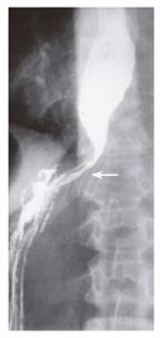
Figure 1 Barium swallow demonstrating smooth, symmetric tapering of the esophagus (arrow), suggesting achalasia.
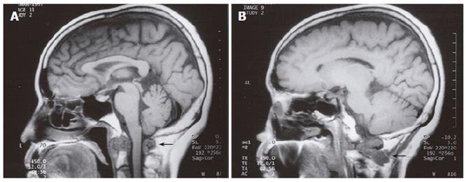
Figure 2 Saggital T1-weighted MRI demonstrating low-signal consistent with marrow replacement at both C1 and the posterior vertebral body of C3 (arrow) (A), and low-signal soft tissue mass adjacent to the C2 vertebral body (arrow) (B).
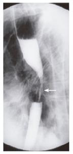
Figure 3 Barium swallow demonstrating irregular stenosis (arrow) involving the middle esophagus.
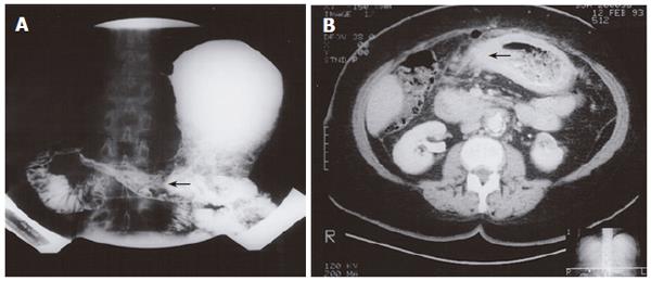
Figure 4 Double-contrast upper GI examination demonstrating irregular narrowing of the gastric antrum (arrow) (A), CT demonstrating gastric mural thickening, perigastric stranding, (arrow) and lymphadenopathy (B).
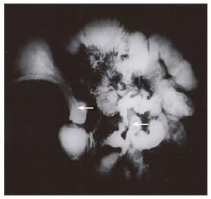
Figure 5 Small bowel enteroclysis showing persistent narrowing of the gastric body and antrum (arrow), prominent irregular gastric rugae with diffuse mucosal serration, and strictures of the ileum.
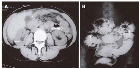
Figure 6 CT demonstrating a dilated loop of small bowel (arrow) and ascites (A), double-contrast barium enema demonstrating fixed eccentric strictures of the ascending colon and splenic flexure (arrows) with a third obscuring lesion seen within the sigmoid colon but not projected in profile on this image (B).
- Citation: Nazareno J, Taves D, Preiksaitis HG. Metastatic breast cancer to the gastrointestinal tract: A case series and review of the literature. World J Gastroenterol 2006; 12(38): 6219-6224
- URL: https://www.wjgnet.com/1007-9327/full/v12/i38/6219.htm
- DOI: https://dx.doi.org/10.3748/wjg.v12.i38.6219


