Copyright
©2006 Baishideng Publishing Group Co.
World J Gastroenterol. Oct 7, 2006; 12(37): 6074-6076
Published online Oct 7, 2006. doi: 10.3748/wjg.v12.i37.6074
Published online Oct 7, 2006. doi: 10.3748/wjg.v12.i37.6074
Figure 1 An upright plain abdominal radiographic imaging revealing 30-40 overt dense opacities in lumen of colonic segments, with oval and well shaped contours, each approximately 1 cm x 1 cm in size.
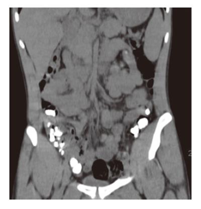
Figure 2 CT showing dense opacities in lumen of colonic segments, with oval and well shaped contours.
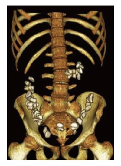
Figure 3 A multiplanar reconstruction and three-dimensional image combined with sectional screening showing all pebbles had passed completely into the colon and no foreign bodies were remained in the ileal segments.
- Citation: Eryilmaz M, Ozturk O, Mentes O, Soylu K, Durusu M, Oner K. Intracolonic multiple pebbles in young adults: Radiographic imaging and conventional approach to a case. World J Gastroenterol 2006; 12(37): 6074-6076
- URL: https://www.wjgnet.com/1007-9327/full/v12/i37/6074.htm
- DOI: https://dx.doi.org/10.3748/wjg.v12.i37.6074


