Copyright
©2006 Baishideng Publishing Group Co.
World J Gastroenterol. Sep 14, 2006; 12(34): 5582-5586
Published online Sep 14, 2006. doi: 10.3748/wjg.v12.i34.5582
Published online Sep 14, 2006. doi: 10.3748/wjg.v12.i34.5582
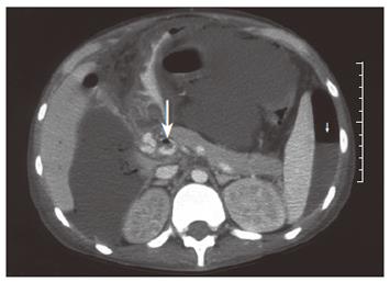
Figure 1 Computed tomography of the abdomen showing evidence of portal venous gas (long arrow), portal vein thrombosis, gross ascites, and pneumoperitoneum (short arrow).
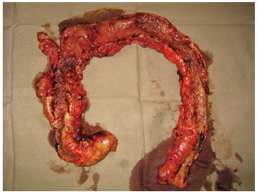
Figure 2 Photograph of the total colectomy specimen showing the classical features of Crohn’s colitis: cobblestone mucosa with skipped lesions.
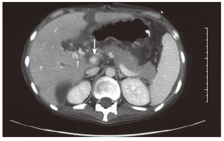
Figure 3 Computed tomography of the abdomen showing evidence of partial recanalization of the portal vein (long arrow) with increasing surrounding collaterals (short arrow).
- Citation: Ng SSM, Yiu RYC, Lee JFY, Li JCM, Leung KL. Portal venous gas and thrombosis in a Chinese patient with fulminant Crohn’s colitis: A case report with literature review. World J Gastroenterol 2006; 12(34): 5582-5586
- URL: https://www.wjgnet.com/1007-9327/full/v12/i34/5582.htm
- DOI: https://dx.doi.org/10.3748/wjg.v12.i34.5582


