Copyright
©2006 Baishideng Publishing Group Co.
World J Gastroenterol. Sep 14, 2006; 12(34): 5569-5572
Published online Sep 14, 2006. doi: 10.3748/wjg.v12.i34.5569
Published online Sep 14, 2006. doi: 10.3748/wjg.v12.i34.5569
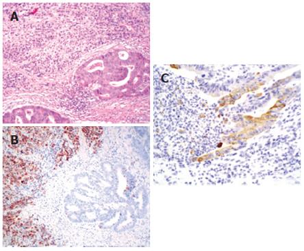
Figure 1 Histological features of rectal tumor showing the interface of a tubular adeonicarcinoma component of primary rectal cancer (right) and a poorly-differentiated adenocarcinoma component metastasized from the stomach (left) (hematoxylin and eosin; x 40) (A), immunohistochemical analysis showing positive MUC2 in metastatic gastric carcinoma and negative MUC2 in primary rectal carcinoma (x 200) (B), and negative CK7 in metastatic gastric carcinoma and positive CK7 in primary rectal carcinoma (x 200) (C).
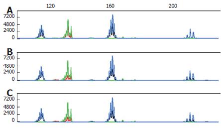
Figure 2 Microsatellite instability phenotype analysis showing no microsatellite instability in the primary rectal tumor (A), the metastatic gastric tumor (B) and the primary gastric tumor (C).
Blue and green line: Normal tissue; Black and red line: Tumor tissue.
- Citation: Roh YH, Lee HW, Kim MC, Lee KW, Roh MS. Collision tumor of the rectum: A case report of metastatic gastric adenocarcinoma plus primary rectal adenocarcinoma. World J Gastroenterol 2006; 12(34): 5569-5572
- URL: https://www.wjgnet.com/1007-9327/full/v12/i34/5569.htm
- DOI: https://dx.doi.org/10.3748/wjg.v12.i34.5569


