Copyright
©2006 Baishideng Publishing Group Co.
World J Gastroenterol. Sep 7, 2006; 12(33): 5408-5411
Published online Sep 7, 2006. doi: 10.3748/wjg.v12.i33.5408
Published online Sep 7, 2006. doi: 10.3748/wjg.v12.i33.5408
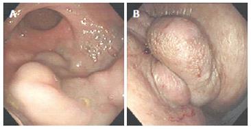
Figure 1 Flexible sigmoidoscopy reveals huge tortuous rectal varices extending from outside the anus to the recto-sigmoid junction (A: rectum, B: outside the anus).
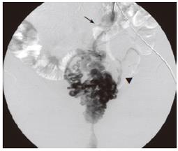
Figure 2 Percutaneous transhepatic portography demonstrates that the rectal varices are supplied by the inferior mesenteric vein (arrow) and flowed into the bilateral internal iliac vein (arrowhead).
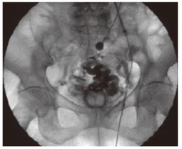
Figure 3 The sclerosant stagnates in the varices as a result of occlusion of the blood flow of the inferior mesenteric vein.
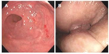
Figure 4 Sigmoidoscopy reveals disappearance of huge tortuous rectal varices extending from outside the anus to the recto-sigmoid junction (A: rectum; B: outside the anus).
- Citation: Okazaki H, Higuchi K, Shiba M, Nakamura S, Wada T, Yamamori K, Machida A, Kadouchi K, Tamori A, Tominaga K, Watanabe T, Fujiwara Y, Nakamura K, Arakawa T. Successful treatment of giant rectal varices by modified percutaneous transhepatic obliteration with sclerosant: Report of a case. World J Gastroenterol 2006; 12(33): 5408-5411
- URL: https://www.wjgnet.com/1007-9327/full/v12/i33/5408.htm
- DOI: https://dx.doi.org/10.3748/wjg.v12.i33.5408


