Copyright
©2006 Baishideng Publishing Group Co.
World J Gastroenterol. Sep 7, 2006; 12(33): 5281-5286
Published online Sep 7, 2006. doi: 10.3748/wjg.v12.i33.5281
Published online Sep 7, 2006. doi: 10.3748/wjg.v12.i33.5281
Figure 1 PCR results of hMLH3 fragments on 2% agarose gel electrophoresis.
M: Molecular marker.
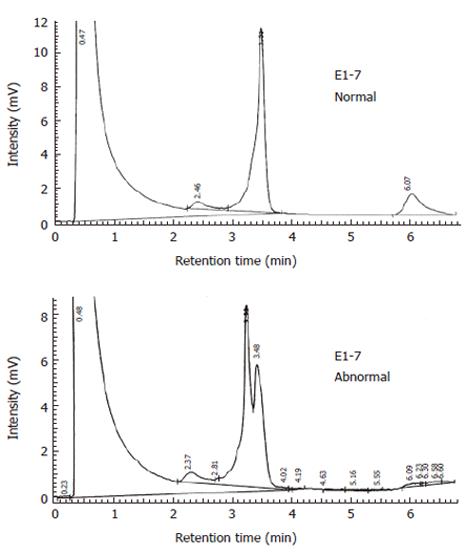
Figure 2 Normal and abnormal DHPLC chromatogram.
The normal control appears as a clear elution peak, while one or more additional earlier eluting peaks result from heteroduplex for mutated DNA.
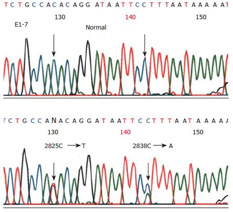
Figure 3 Sequencing results revealing two heterozygous variants in fragment 7 of exon 1, C2825T and C2838A.
On the basis of amino acid variation, C2825T (Thr9421Ile) is considered a missense mutation, while C2838A (Ser947Ser) a polymorphism.
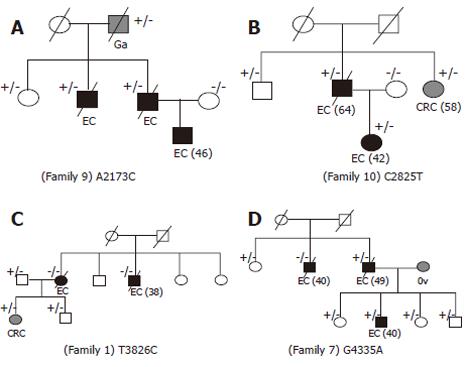
Figure 4 Pedigree of families with hMLH3 variants.
Symbols and abbreviations used are denoted as black symbols for esophageal cancer, gray symbols for other cancers; EC: Esophageal cancer; CRC: Colorectal cancer; Ga: Gastric cancer; Ov: Ovarian cancer; Numbers next to the diagnosis denote age at onset; genotypes are on the top right of family member symbols; +: Variant carrier; -: Nonvariant carrier.
- Citation: Liu HX, Li Y, Jiang XD, Yin HN, Zhang L, Wang Y, Yang J. Mutation screening of mismatch repair gene Mlh3 in familial esophageal cancer. World J Gastroenterol 2006; 12(33): 5281-5286
- URL: https://www.wjgnet.com/1007-9327/full/v12/i33/5281.htm
- DOI: https://dx.doi.org/10.3748/wjg.v12.i33.5281


