Copyright
©2006 Baishideng Publishing Group Co.
World J Gastroenterol. Aug 21, 2006; 12(31): 5087-5090
Published online Aug 21, 2006. doi: 10.3748/wjg.v12.i31.5087
Published online Aug 21, 2006. doi: 10.3748/wjg.v12.i31.5087
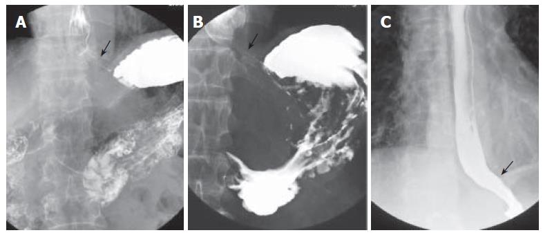
Figure 1 A: Barium esophagography and upper gastrointestinal series showing an intact gastroesophageal junction; B: dilated distal esophageal lumen with food retention and narrowing of gastroesophageal junction after truncal vagotomy; C: barium esophagography showing a non-dilated esophageal lumen with smooth passage of contrast medium through gastroesophageal junction after pneumodilations.
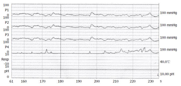
Figure 2 Confirmation of achalasia by manometric findings of esophageal aperistalsis and incomplete relaxation of the lower esophageal sphincter during wet swallowing.
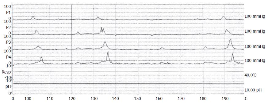
Figure 3 Manomteric studies showing restoration of esophageal motility with normal peristalsis as a healthy subject after dilations.
- Citation: Chuah SK, Kuo CM, Wu KL, Changchien CS, Hu TH, Wang CC, Chiu YC, Chou YP, Hsu PI, Chiu KW, Kuo CH, Chiou SS, Lee CM. Pseudoachalasia in a patient after truncal vagotomy surgery successfully treated by subsequent pneumatic dilations. World J Gastroenterol 2006; 12(31): 5087-5090
- URL: https://www.wjgnet.com/1007-9327/full/v12/i31/5087.htm
- DOI: https://dx.doi.org/10.3748/wjg.v12.i31.5087


