Copyright
©2006 Baishideng Publishing Group Co.
World J Gastroenterol. Aug 21, 2006; 12(31): 5081-5083
Published online Aug 21, 2006. doi: 10.3748/wjg.v12.i31.5081
Published online Aug 21, 2006. doi: 10.3748/wjg.v12.i31.5081
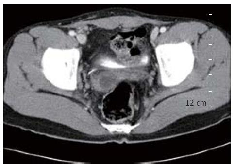
Figure 1 Abdominal computed tomography revealing an oval-shaped cyst with a thickened wall in anterolateral to the rectum.
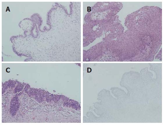
Figure 2 Microscopically, the cyst wall showing a variety of lining epithelia including tall columnar or cuboidal (A), squamous (B), and transitional (C) epithelium (HE, x 200), the underlying stroma showing a mild infiltrate of chronic inflammatory cells, immunohistochemical staining showing negative prostate specific antigen in the lining of epithelial cells (D) (PAP, x 200).
- Citation: Jang SH, Jang KS, Song YS, Min KW, Han HX, Lee KG, Paik SS. Unusual prerectal location of a tailgut cyst: A case report. World J Gastroenterol 2006; 12(31): 5081-5083
- URL: https://www.wjgnet.com/1007-9327/full/v12/i31/5081.htm
- DOI: https://dx.doi.org/10.3748/wjg.v12.i31.5081


