Copyright
©2006 Baishideng Publishing Group Co.
World J Gastroenterol. Aug 14, 2006; 12(30): 4927-4929
Published online Aug 14, 2006. doi: 10.3748/wjg.v12.i30.4927
Published online Aug 14, 2006. doi: 10.3748/wjg.v12.i30.4927
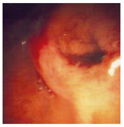
Figure 1 Endoscopic view showing a dilated papillary orifice exiting mucus.
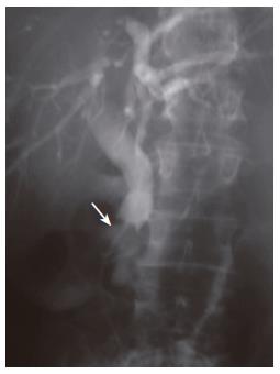
Figure 2 ERCP demonstrating an intraluminal filling defect in the distal common bile duct.
A 8.5Fr stent has been placed.
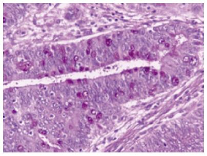
Figure 3 Surgical specimen showing that the tumor consists of tall, columnar, pseudostratified epithelial cells with elongated or round atypical nuclei.
Some of the cells contain large amounts of mucin within the apical cytoplasm (PAS X 400).
- Citation: Katsinelos P, Basdanis G, Chatzimavroudis G, Karagiannoulou G, Katsinelos T, Paroutoglou G, Papaziogas B, Paraskevas G. Pancreatitis complicating mucin-hypersecreting common bile duct adenoma. World J Gastroenterol 2006; 12(30): 4927-4929
- URL: https://www.wjgnet.com/1007-9327/full/v12/i30/4927.htm
- DOI: https://dx.doi.org/10.3748/wjg.v12.i30.4927


