Copyright
©2006 Baishideng Publishing Group Co.
World J Gastroenterol. Aug 14, 2006; 12(30): 4918-4921
Published online Aug 14, 2006. doi: 10.3748/wjg.v12.i30.4918
Published online Aug 14, 2006. doi: 10.3748/wjg.v12.i30.4918
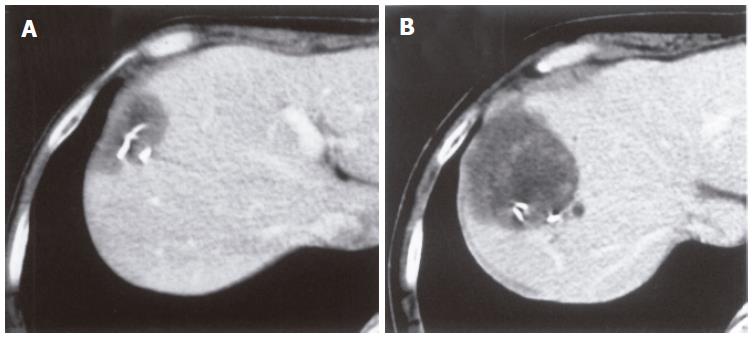
Figure 1 A low-density lesion at the cut surface of the liver in December, 1998 (A) and two years later, the mass increased in size to approximately 5 cm in diameter (B).
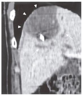
Figure 2 A low-density cystic tumor in S8 with local dilatation of the IHBD (arrows).
The tumor was bordered by the diaphragm (arrow heads).
Figure 3 A low intensity signal on T1-weighted (A) and a high intensity signal on T2-weighted (B) images.
The edge of the tumor was slightly enhanced by gadolinium, while the inside was heterogeneous (C).
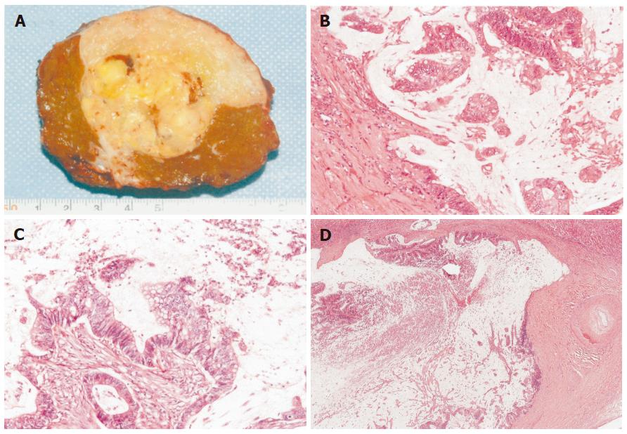
Figure 4 Macroscopic finding (A), well-differentiated mucinous adenocarcinoma (B) similar to cecal cancer (C), extension of tumor cells along the lumen of the biliary ducts with the non-neoplastic epitheliumreplaced (D).
(B) HE, ×100; (C) HE, ×100; (D) HE, ×18.
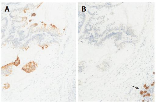
Figure 5 Immunohisotochemically, tumor cells positive for CK 20 (A), but negative for CK7 (B), normal epithelial cells of IHBD positive for CK7 (arrow).
(A) × 100; (B) × 100.
- Citation: Tokai H, Kawashita Y, Eguchi S, Kamohara Y, Takatsuki M, Okudaira S, Tajima Y, Hayashi T, Kanematsu T. A case of mucin producing liver metastases with intrabiliary extension. World J Gastroenterol 2006; 12(30): 4918-4921
- URL: https://www.wjgnet.com/1007-9327/full/v12/i30/4918.htm
- DOI: https://dx.doi.org/10.3748/wjg.v12.i30.4918


