Copyright
©2006 Baishideng Publishing Group Co.
World J Gastroenterol. Aug 14, 2006; 12(30): 4866-4869
Published online Aug 14, 2006. doi: 10.3748/wjg.v12.i30.4866
Published online Aug 14, 2006. doi: 10.3748/wjg.v12.i30.4866
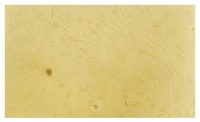
Figure 1 After 3 passages, on the 14th d, the cell showed uniform morphology: fusiform shape or polygon shape, like fibroblast and grew as grass bunch or whirlpool (100 ×).
Figure 2 Growth condition of MSC cultured with cholestatic serum.
A: Cultured with cholestatic serum for 7 d, peripheral cell of the clone showed cuboidal morphology, which is characteristic of hepatocytes (100 ×); B: Cultured with cholestatic serum for 35 d, all MSCs showed cuboidal morphology, which is characteristic of hepatocytes (100 ×).
Figure 3 Growth condition of MSC cultured with HGF.
A: Cultured with HGF for 7 d, peripheral cell of the clone showed cuboidal morphology, which is characteristic of hepatocytes(100×); B: Cultured with HGF for 35 d, all MSCs showed cuboidal morphology, which is characteristic of hepatocytes (100 ×).
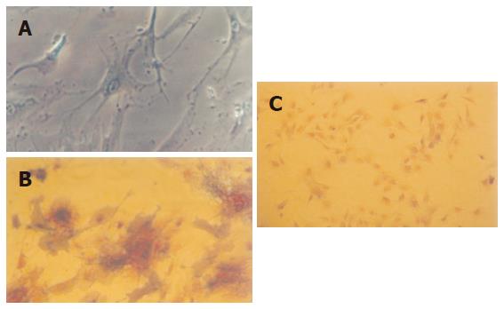
Figure 4 MSC identification.
A: Induced by vitamin C, dexamethasone and β sodium glycerophosphate, MSC differentiated into osteoblast (200×); B: Alkaline phosphatase stain showed positive (400 ×); C: Alkaline phosphatase stain negative control (100 ×).
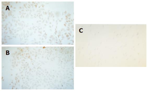
Figure 5 Expression of CK18 and AFP.
A: Cultured with cholestatic serum for 14 d, MSC expressed AFP by S-P method (100×); B: Cultured with HGF for 14 d, MSC expressed CK18 by S-P method (100 ×); C: Negative control (100 ×).
Figure 6 Expression of albumin.
A: Culture with cholestatic serum for 35 d, expression of albumin was confirmed by in situ hybridization (FITC dyeing ) (400 ×); B: Culture with HGF for 35 d, expression of albumin was confirmed by in situ hybridization (FITC dyeing ) (400 ×).
- Citation: Li W, Liu SN, Luo DD, Zhao L, Zeng LL, Zhang SL, Li SL. Differentiation of hepatocytoid cell induced from whole-bone-marrow method isolated rat myeloid mesenchymal stem cells. World J Gastroenterol 2006; 12(30): 4866-4869
- URL: https://www.wjgnet.com/1007-9327/full/v12/i30/4866.htm
- DOI: https://dx.doi.org/10.3748/wjg.v12.i30.4866


