Copyright
©2006 Baishideng Publishing Group Co.
World J Gastroenterol. Jan 21, 2006; 12(3): 431-436
Published online Jan 21, 2006. doi: 10.3748/wjg.v12.i3.431
Published online Jan 21, 2006. doi: 10.3748/wjg.v12.i3.431
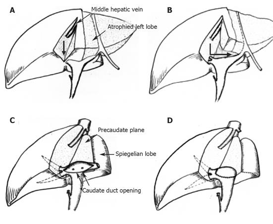
Figure 1 Schematic drawing of VHE procedure during left lobectomy.
A: Hepatotomy commences vertically from the Cantlie’s line to the direction of the hilar bile duct (thick arrow); B: after reaching the hilum, the hepatotomy is continued along the pre-caudate plane (angled thick arrow); C: the ventral portion of the hilar hepatic duct is opened along its direction; D: after completion of the necessary ductal procedure.
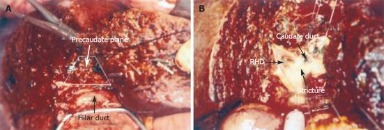
Figure 2 A: Dissecting along the pre-caudate plane allows the ventral portion of the hilar duct to be exposed during left lobectomy; B: the hilar duct was opened along its direction; RHD: right hepatic duct opening.
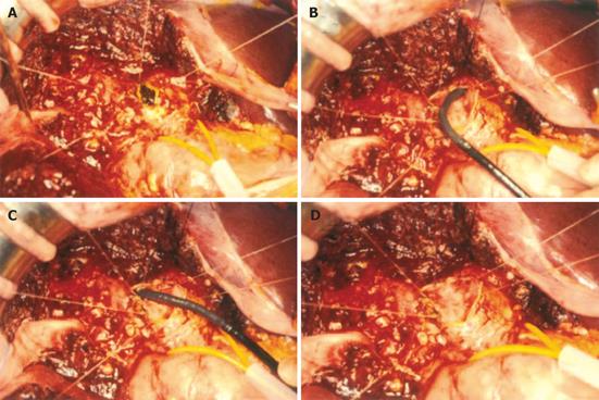
Figure 3 During central lobectomy, the hilar duct was exposed and opened.
A: Extraction of the IHD stones; B and C: application of intra-operative choledochoscope to the left and right IHD; D: after the removal of all intra-hepatic stones.
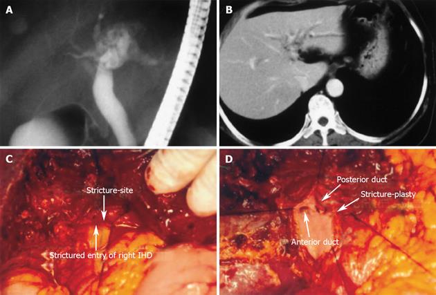
Figure 4 A and B: Pre-operative imaging of hepatolithiasis with hilar duct stricture; C: opened hilar IHD shows narrow entry of right IHD (arrow); D: after stricture-plasty both entries of the right anterior and posterior ducts are seen.
- Citation: Kim BW, Wang HJ, Kim WH, Kim MW. Favorable outcomes of hilar duct oriented hepatic resection for high grade Tsunoda type hepatolithiasis. World J Gastroenterol 2006; 12(3): 431-436
- URL: https://www.wjgnet.com/1007-9327/full/v12/i3/431.htm
- DOI: https://dx.doi.org/10.3748/wjg.v12.i3.431


