Copyright
©2006 Baishideng Publishing Group Co.
World J Gastroenterol. Jul 28, 2006; 12(28): 4602-4603
Published online Jul 28, 2006. doi: 10.3748/wjg.v12.i28.4602
Published online Jul 28, 2006. doi: 10.3748/wjg.v12.i28.4602
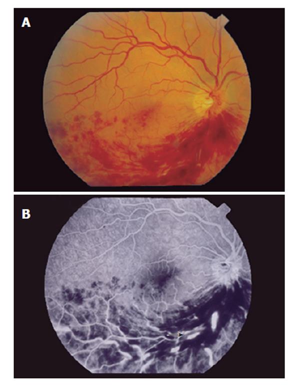
Figure 1 A: Fundus photograph of the right eye with extensive intraretinal hemorrhages and cotton-wool spots in the inferonasal and inferotemporal regions; B: Fluorescein angiography with segmental hypoperfusion, dilation and tortuosity of the retinal veins (arrow), compatible with inferior branch retinal vein thrombosis.
- Citation: Gonçalves LL, Farias AQ, Gonçalves PL, D’Amico EA, Carrilho FJ. Branch retinal vein thrombosis and visual loss probably associated with pegylated interferon therapy of chronic hepatitis C. World J Gastroenterol 2006; 12(28): 4602-4603
- URL: https://www.wjgnet.com/1007-9327/full/v12/i28/4602.htm
- DOI: https://dx.doi.org/10.3748/wjg.v12.i28.4602


