Copyright
©2006 Baishideng Publishing Group Co.
World J Gastroenterol. Jul 28, 2006; 12(28): 4498-4503
Published online Jul 28, 2006. doi: 10.3748/wjg.v12.i28.4498
Published online Jul 28, 2006. doi: 10.3748/wjg.v12.i28.4498
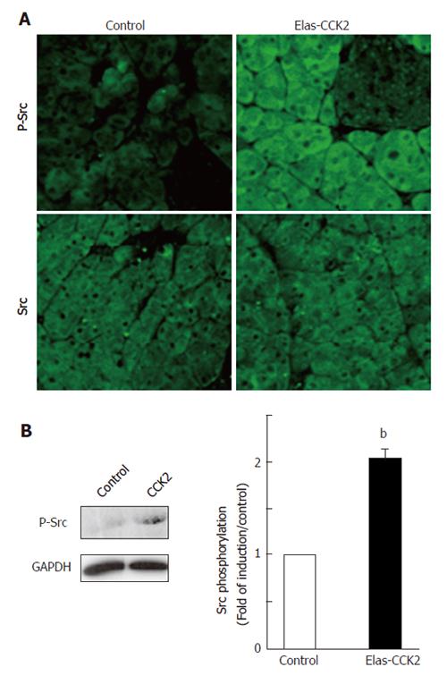
Figure 1 CCK2R expression in acini of Elas-CCK2 mice induced the activation of Src.
Immunohistochemistry analysis on paraffin-embedded pancreatic tissues or western-blots on lysates from isolated acinar cells were performed using antibodies specific for total Src (SRC) or detecting the activated form of the protein, PY418-Src (P-Src) as indicated. Representative data from 3 experiments (3 different animals in each group) are shown. A: Original magnification: 40 X; B: Blots were also probed with an antibody against GAPDH to ensure equal loading of proteins. Results of western-blots quantification are presented as mean ± SE. bP < 0.01 vs control.
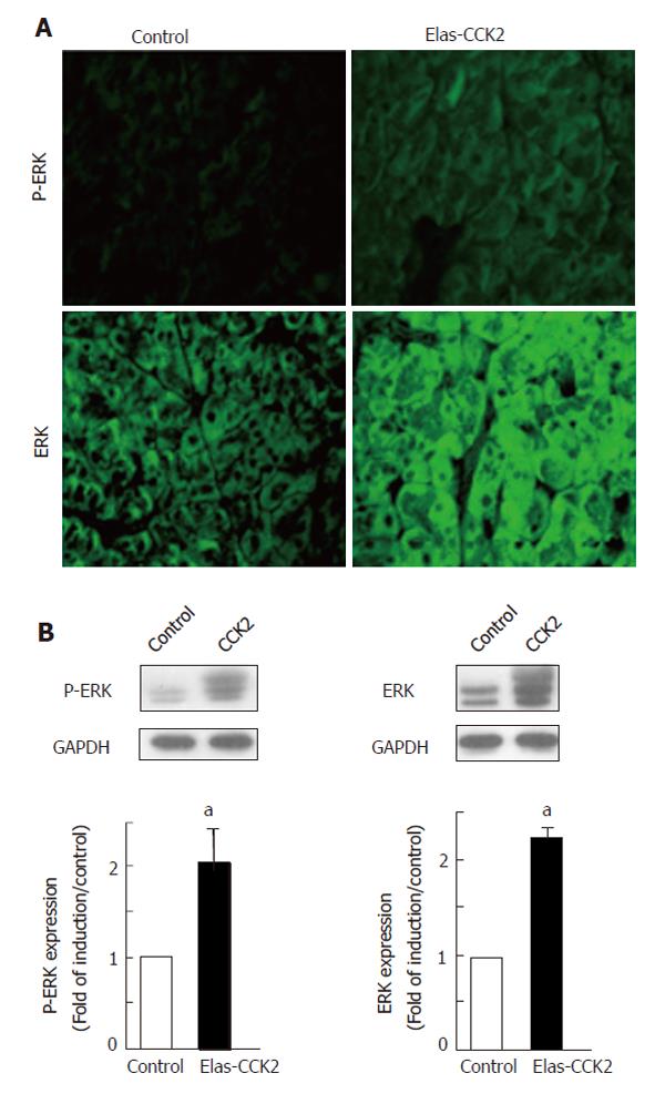
Figure 2 CCK2R expression in acini of Elas-CCK2 mice induces the activation of the ERK pathway.
Immunohistochemistry analysis on paraffin-embedded pancreatic tissues or western-blots on lysates from isolated acinar cells were performed using antibodies specific for total ERK (ERK) or detecting the activated form of the protein, Phospho-ERK (P-ERK) as indicated. Representative data from 3 experiments (3 different animals in each group) are shown. A: Original magnification: 40 X; B: Blots were also probed with an antibody against GAPDH to ensure equal loading of proteins. Results of western-blots quantification are presented as mean ± SE. aP < 0.05 vs control.
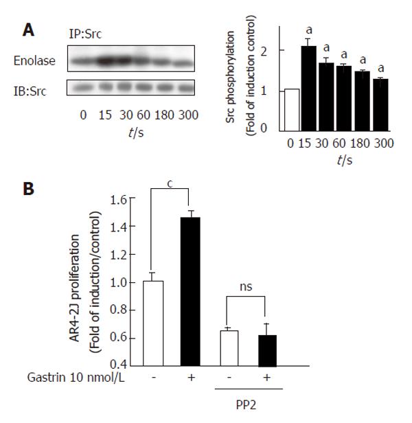
Figure 3 Role of Src in proliferation of tumour pancreatic acinar cells induced by the CCK2R.
AR4-2J cells were stimulated with Gastrin (10 nmol/L) for the times indicated (A) or 15 s (B). A: Src activity was determined as described in Methods. Immunoprecipitated proteins were also analysed by western-blot using anti-Src antibodies. Results of autoradiography quantification are presented as mean ± SE. aP < 0.05 vs control. B: Serum-starved AR4-2J cells were treated with Gastrin for 48 h in the presence or absence of PP2 (10 μmol/L), and the proliferation determined as described in Methods. Data are presented as mean ± SE. cP < 0.001 vs control.
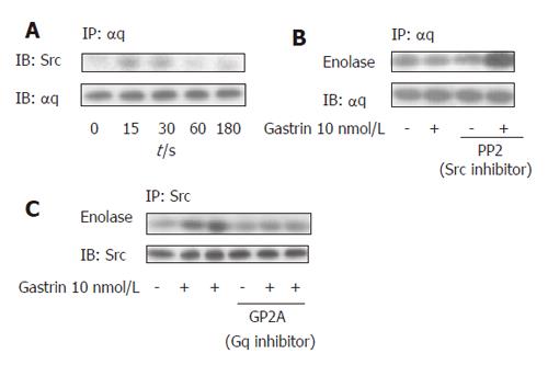
Figure 4 Src activation by the CCK2R in tumour pancreatic acinar cells (A-C).
AR4-2J cells were stimulated with Gastrin (10 nmol/L) for the times indicated (A) or for 15 s (B, C). When indicated, cells where pretreated with 10 μmol/L of PP2 or GP2A for 30 min. A: Cell lysates were immunoprecipitated (IP) with an anti-αq antibody and immunoblotted (IB) with the anti-Src antibody. The blots were also probed with the antibody used for immunoprecipitation to ensure equal loading of proteins; B, C: following immunoprecipitation with an anti-αq or an anti-Src antibody, Src activity was determined as described in Methods. Immunoprecipitated proteins were also analyzed by western blot using the anti-αq or anti-Src antibodies as indicated.
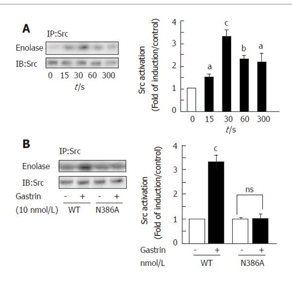
Figure 5 Involvement of the NPXXY motif in Src activation by the CCK2R.
A: COS-7 cells transfected with the human CCK2R were stimulated with Gastrin (10 nmol/L) for the time indicated. Src kinase activity was determined as described in Methods. Immunoprecipitated proteins were also analyzed by western-blot using anti-Src antibodies. Results of autoradiography quantification are presented as mean ± SE. aP < 0.05 vs control, bP < 0.01 vs control, cP < 0.001 vs control; B: COS-7 cells transfected with the WT or N386A mutant CCK2R were stimulated or not with Gastrin (10 nmol/L) for 30 s and Src kinase activity determined as described in Methods. Immunoprecipitated proteins were also analysed by western-blot using anti-Src antibodies. Results of autoradiography quantification are presented as mean ± SE. cP < 0.001 vs control.
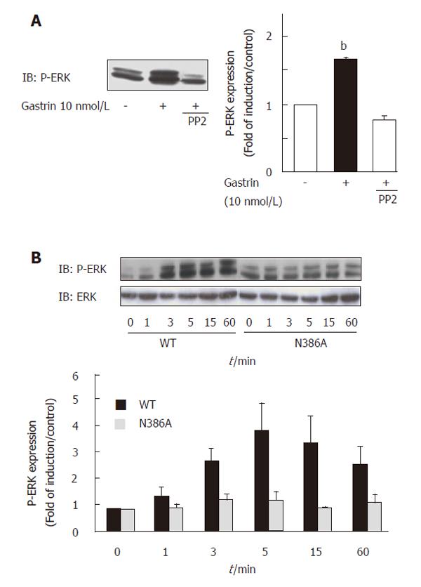
Figure 6 Involvement of the NPXXY motif in ERK activation by the CCK2R.
COS-7 cells transfected with the WT or N386A mutant CCK2R were stimulated or not with Gastrin (10 nmol/L) for 5 min (A) or the time indicated (B). When indicated, cells were pretreated with the Src inhibitor PP2 (10 μmol/L). Equal amounts of protein were analyzed by western-blot using anti-phospho-ERK antibodies. Blots were also reprobed with antibodies directed against total ERK proteins. Results of autoradiography quantification are presented as mean ± SE. bP < 0.01 vs control.
- Citation: Ferrand A, Vatinel S, Kowalski-Chauvel A, Bertrand C, Escrieut C, Fourmy D, Dufresne M, Seva C. Mechanism for Src activation by the CCK2 receptor: Patho-physiological functions of this receptor in pancreas. World J Gastroenterol 2006; 12(28): 4498-4503
- URL: https://www.wjgnet.com/1007-9327/full/v12/i28/4498.htm
- DOI: https://dx.doi.org/10.3748/wjg.v12.i28.4498


