Copyright
©2006 Baishideng Publishing Group Co.
World J Gastroenterol. Jul 14, 2006; 12(26): 4264-4266
Published online Jul 14, 2006. doi: 10.3748/wjg.v12.i26.4264
Published online Jul 14, 2006. doi: 10.3748/wjg.v12.i26.4264
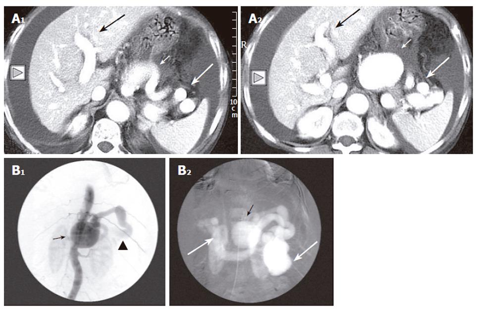
Figure 1 A: Post contrast CT scan reveals a tortuous and aneurysmatic splenic artery of maximum diameter of approximately 52 mm (short white arrow) associated with dilated vessels at the splenic hilum (long white arrow) and early opacification of the portal axis (long black arrow).
In addition ascites is present as well (small triangle); B: Celiac angiogram confirms the presence of the splenic artery aneurysm (black arrow) in contiguity with the markedly dilated splenic vein and the premature and intense filling of the splenoportal trunk (black arrowhead and white arrows).
Figure 2 Selective transarterial catheterization (long black arrow) and embolization of the aneurysmal sac with numerous adequate metallic macrocoils (small black arrows) resulted in full occlusion of the sac and the fistulous tract enabling thus the reduction of the pressure in the splenoportal circulation.
- Citation: Siablis D, Papathanassiou ZG, Karnabatidis D, Christeas N, Katsanos K, Vagianos C. Splenic arteriovenous fistula and sudden onset of portal hypertension as complications of a ruptured splenic artery aneurysm: Successful treatment with transcatheter arterial embolization. A case study and review of the literature. World J Gastroenterol 2006; 12(26): 4264-4266
- URL: https://www.wjgnet.com/1007-9327/full/v12/i26/4264.htm
- DOI: https://dx.doi.org/10.3748/wjg.v12.i26.4264


