Copyright
©2006 Baishideng Publishing Group Co.
World J Gastroenterol. Jul 14, 2006; 12(26): 4175-4178
Published online Jul 14, 2006. doi: 10.3748/wjg.v12.i26.4175
Published online Jul 14, 2006. doi: 10.3748/wjg.v12.i26.4175
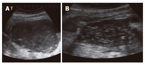
Figure 1 EUS images of an infected pancreatic pseudocyst (abscess) prior (A) and after placement (B) of an external drainage.
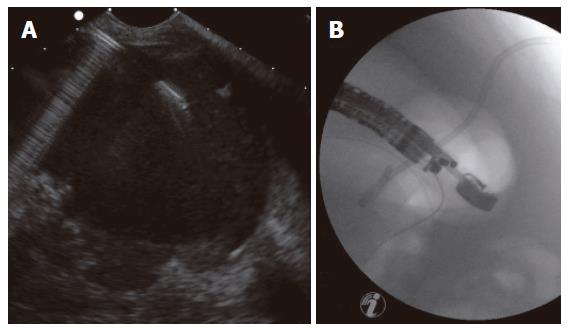
Figure 2 EUS-guided drainage of a pancreatic pseudocyst after previous external drainage: (A) EUS image; (B) Subsequent fluoroscopy control image of correct placement of the drainage.
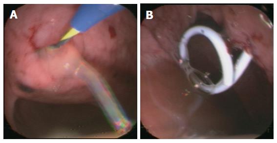
Figure 3 Endoscopy-guided cystogastrostomy via guide wire (A) and placement of an internal drainage (B).
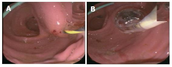
Figure 4 Endoscopic images: (A) EUS-guided puncture after previous external drainage; (B) Balloon dilatation via guide wire.
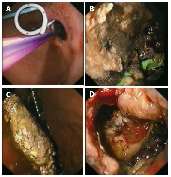
Figure 5 Endoscopic view: (A) After EUS-guided placement of an internal drainage (external drainage previously); (B) Into the pseudocystic cavity showing necroses; (C) Showing necrosectomy; (D) Through the transgastrocystic opening into the necrosectomized pseudocystic cavity.
- Citation: Will U, Wegener C, Graf KI, Wanzar I, Manger T, Meyer F. Differential treatment and early outcome in the interventional endoscopic management of pancreatic pseudocysts in 27 patients. World J Gastroenterol 2006; 12(26): 4175-4178
- URL: https://www.wjgnet.com/1007-9327/full/v12/i26/4175.htm
- DOI: https://dx.doi.org/10.3748/wjg.v12.i26.4175


