Copyright
©2006 Baishideng Publishing Group Co.
World J Gastroenterol. Jul 14, 2006; 12(26): 4156-4160
Published online Jul 14, 2006. doi: 10.3748/wjg.v12.i26.4156
Published online Jul 14, 2006. doi: 10.3748/wjg.v12.i26.4156
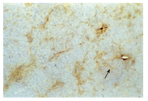
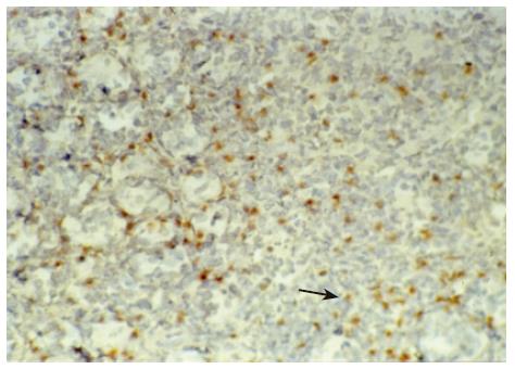
Figure 2 RANTES immunohistochemical staining of pancreas allografts.
Mild expression was observed at 1 d post transplantation (× 200).
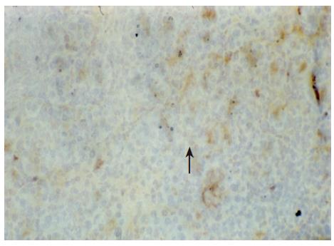
Figure 3 IP-10 immunohistochemical staining of pancreas allografts.
Moderate expression was observed at 4 d post transplantation (× 200).
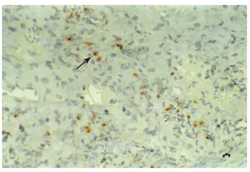
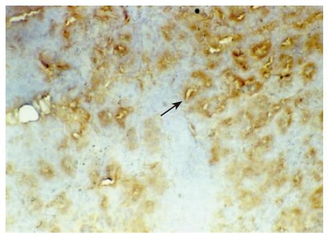
Figure 1 IP-10 immunohistochemical staining of pancreas allografts.
Mild expression was observed at 1 d post transplantation (× 200).
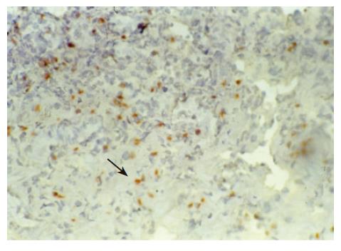
Figure 4 RANTES immunohistochemical staining of pancreas allografts.
Moderate expression was observed at 4 d post transplantation (× 200).
Figure 5 IP-10 immunohistochemical staining of pancreas allografts.
Strong positive expression was observed at 7 d post transplantation (× 200).
Figure 6 RANTES immunohistochemical staining of pancreas allografts.
Strong positive expression was observed at 7 d post transplantation (× 200).
- Citation: Zhu J, Xu ZK, Miao Y, Liu XL, Zhang H. Changes of inducible protein-10 and regulated upon activation, normal T cell expressed and secreted protein in acute rejection of pancreas transplantation in rats. World J Gastroenterol 2006; 12(26): 4156-4160
- URL: https://www.wjgnet.com/1007-9327/full/v12/i26/4156.htm
- DOI: https://dx.doi.org/10.3748/wjg.v12.i26.4156


