Copyright
©2006 Baishideng Publishing Group Co.
World J Gastroenterol. Jul 7, 2006; 12(25): 4106-4108
Published online Jul 7, 2006. doi: 10.3748/wjg.v12.i25.4106
Published online Jul 7, 2006. doi: 10.3748/wjg.v12.i25.4106
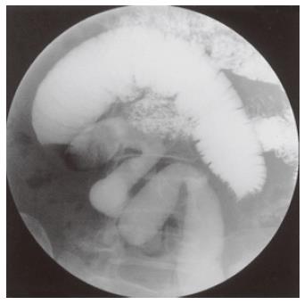
Figure 1 UGI series showing a narrow segment of the small intestine with the disappearance of Kerckring’s folds.
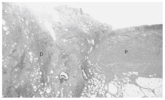
Figure 2 Resected specimens revealed histologically a complete destruction of the jejunal wall (D).
P: Preserved wall of the intestine with an infiltration of inflammatory cells.
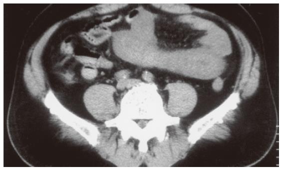
Figure 3 CT showing a remarkable thickening of the intestinal wall.
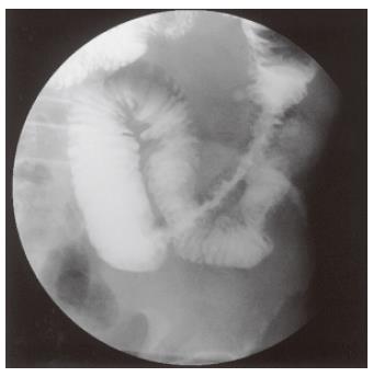
Figure 4 UGI series showing a narrow segment of the small intestine with the preservation of Kerckring’s folds.
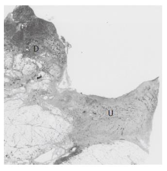
Figure 5 Resected speci-mens revealed histologically a perforated ulceration (U) with an infiltration of inflammatory cells.
D: Destructive wall of the intestine.
- Citation: Matsuo S, Azuma T, Susumu S, Yamaguchi S, Obata S, Hayashi T. Small bowel anisakiosis: A report of two cases. World J Gastroenterol 2006; 12(25): 4106-4108
- URL: https://www.wjgnet.com/1007-9327/full/v12/i25/4106.htm
- DOI: https://dx.doi.org/10.3748/wjg.v12.i25.4106


