Copyright
©2006 Baishideng Publishing Group Co.
World J Gastroenterol. Jul 7, 2006; 12(25): 4014-4019
Published online Jul 7, 2006. doi: 10.3748/wjg.v12.i25.4014
Published online Jul 7, 2006. doi: 10.3748/wjg.v12.i25.4014
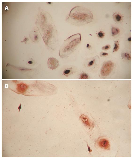
Figure 1 Cells positively stained for AFP (A) and ALB (B) in inducing groups (400 ×).
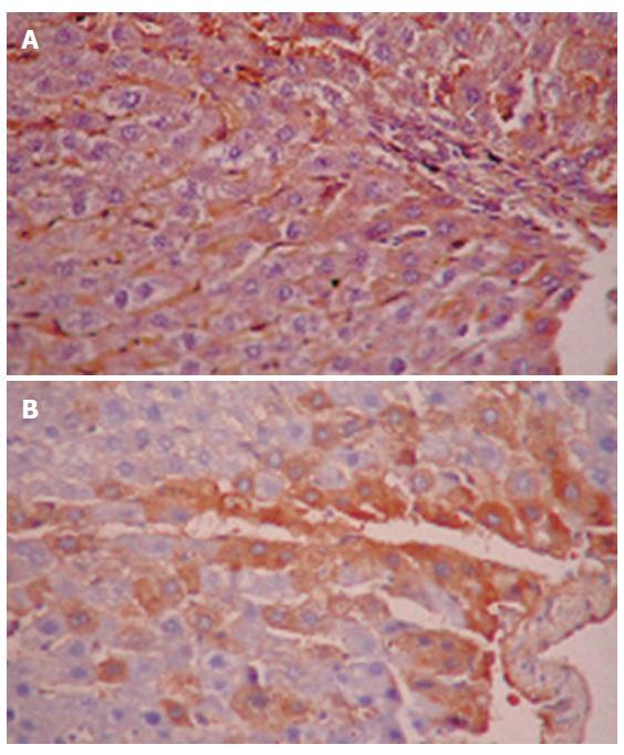
Figure 2 Expression of human ALB (A) and AFP (B) in rat liver tissue (200 ×).
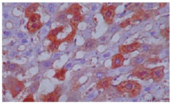
Figure 3 Human ALB positively conjugated nuclear cells in rat liver tissue (400 ×).
Figure 4 PCR-amplified DNA.
M: 100 bp marker; N: Negative control, the products of PCR templated by water; H: Positive control, the products of PCR templated by human fetal liver tissue DNA; 1-5: DNA extracted from rat liver tissue embedded in paraffin in group A; 6-10: DNA extracted from rat liver tissue embedded in paraffin in group B; 11-15: DNA extracted from rat liver tissue embedded in paraffin in group C.
-
Citation: Tang XP, Zhang M, Yang X, Chen LM, Zeng Y. Differentiation of human umbilical cord blood stem cells into hepatocytes
in vivo andin vitro . World J Gastroenterol 2006; 12(25): 4014-4019 - URL: https://www.wjgnet.com/1007-9327/full/v12/i25/4014.htm
- DOI: https://dx.doi.org/10.3748/wjg.v12.i25.4014


