Copyright
©2006 Baishideng Publishing Group Co.
World J Gastroenterol. Jul 7, 2006; 12(25): 3965-3969
Published online Jul 7, 2006. doi: 10.3748/wjg.v12.i25.3965
Published online Jul 7, 2006. doi: 10.3748/wjg.v12.i25.3965
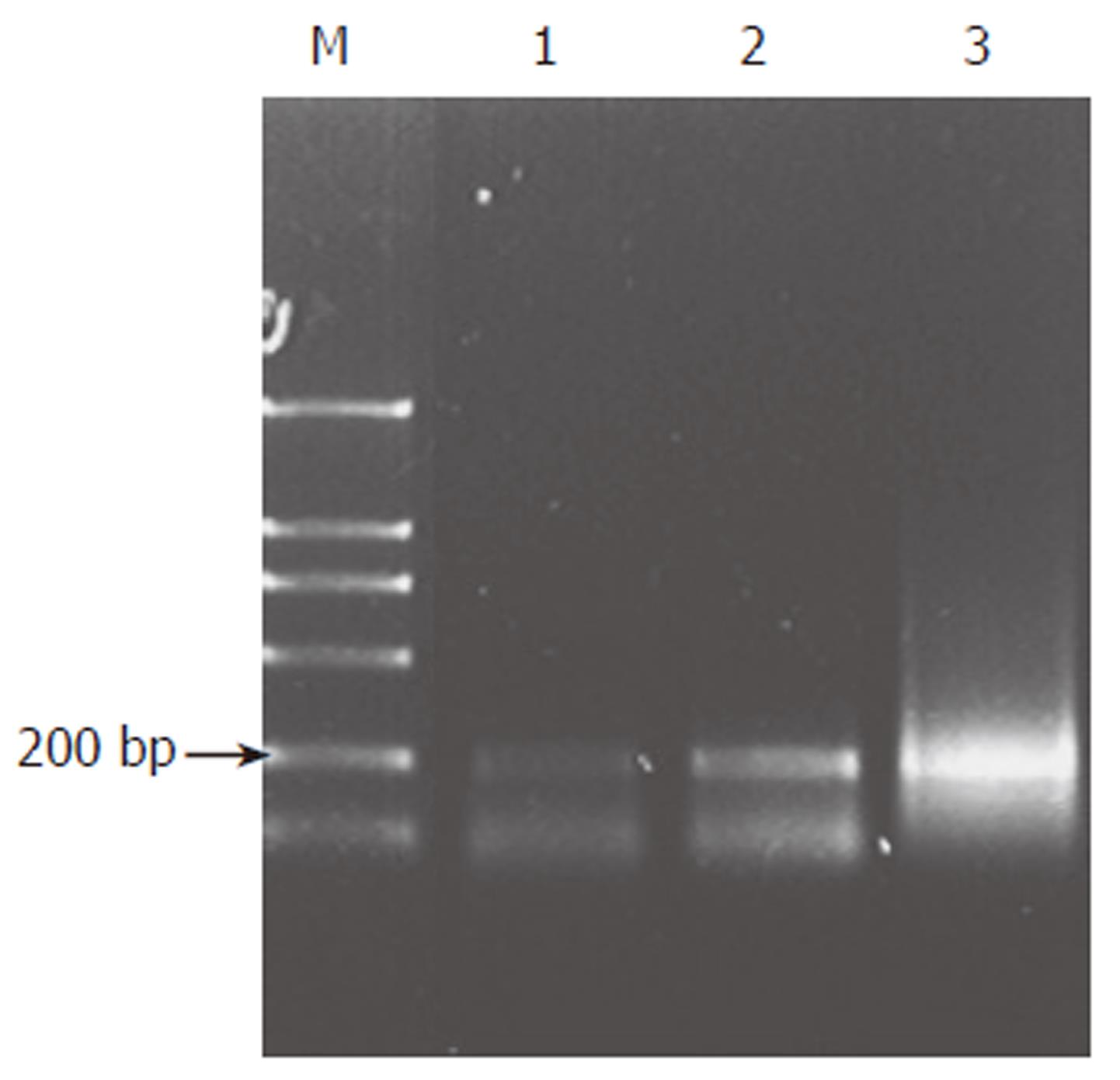
Figure 1 Results of Shh RT-PCR.
M: MarkerDL-2000; 1: Non-cancerous gastric tissues; 2: Well differentiated tubular adenocarcinoma; 3: Poorly differentiated tubular adenocarcinoma.
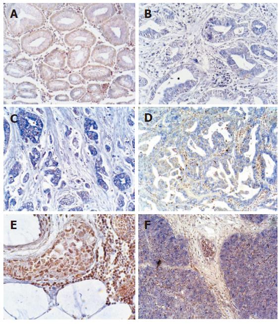
Figure 2 Results of Shh expression by in situ hybridization, blue represents positive.
A: Non-cancerous gastric tissues (× 100); B: Well-moderately differentiated tubular adenocarcinoma (× 100); C: Poorly differentiated tubular adenocarcinoma (× 100); D: Papillary adenocarcinoma (× 100); E: Mucinous adenocarcinoma (× 200); F: Squamous cell carcinoma (× 100).
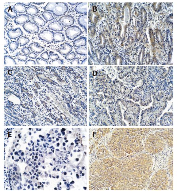
Figure 3 Results of Shh expression by immunohistochemistry (brown represents positive).
A: Non-cancerous gastric tissues (× 100); B: Well-moderately differentiated tubular adenocarcinoma (× 100); C: Poorly differentiated adenocarcinoma (× 100); D: Papillary adenocarcinoma (× 100); E: Mucinous adenocarcinoma (× 200); F: Squamous cell carcinoma (× 100).
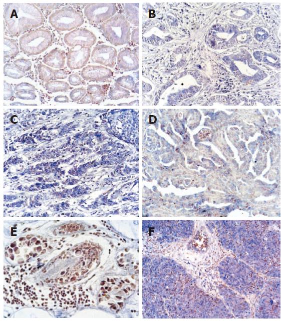
Figure 4 Results of Gli1 by in situ hybridization( blue represents positive).
A: Non-cancerous stomach tissues (× 100); B: Moderately differentiated tubular adenocarcinoma (× 100); C: Poorly differentiated tubular adenocarcinoma (×100); D: Papillary adenocarcinoma (× 100); E: Mucinous adenocarcinoma (× 200); F: Squamous cell carcinoma (× 100).
- Citation: Ma XL, Sun HJ, Wang YS, Huang SH, Xie JW, Zhang HW. Study of Sonic hedgehog signaling pathway related molecules in gastric carcinoma. World J Gastroenterol 2006; 12(25): 3965-3969
- URL: https://www.wjgnet.com/1007-9327/full/v12/i25/3965.htm
- DOI: https://dx.doi.org/10.3748/wjg.v12.i25.3965


