Copyright
©2006 Baishideng Publishing Group Co.
World J Gastroenterol. Jun 28, 2006; 12(24): 3944-3947
Published online Jun 28, 2006. doi: 10.3748/wjg.v12.i24.3944
Published online Jun 28, 2006. doi: 10.3748/wjg.v12.i24.3944
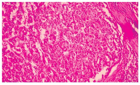
Figure 1 Micrograph of histological section of the neuroendocrine gastric carcinoma showing a proeminent trabecular architecture, organoid pattern with pseudoglandular structure, uniform cells and abundant mitoses.
(Hematoxilin & Eosin staining; original magnification, × 200).
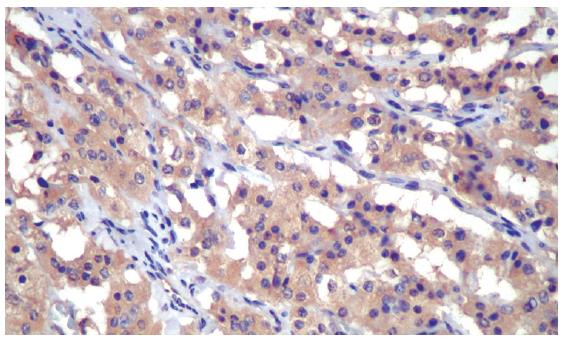
Figure 2 Immunohistochemistry of the neuroendocrine gastric carcinoma showing intense and diffuse somatostatin-positive imunoreactivity in the neoplastic cells (Immunoperoxidase, primary antibody anti-human somatostatin-code A0566-Dako A/S®, Copenhagen, Denmark, original magnification, × 400).
- Citation: Waisberg J, Matos LL, Mader AMAA, Pezzolo S, Eher EM, Capelozzi VL, Speranzini MB. Neuroendocrine gastric carcinoma expressing somatostatin: A highly malignant, rare tumor. World J Gastroenterol 2006; 12(24): 3944-3947
- URL: https://www.wjgnet.com/1007-9327/full/v12/i24/3944.htm
- DOI: https://dx.doi.org/10.3748/wjg.v12.i24.3944


